Jung Minjeong, Lee Jaemeun, Seo Hye-Young, Lim Ji Sun, Kim Eun-Kyoung
Department of Brain Science, Daegu Gyeongbuk Institute of Science & Technology, Daegu, Korea.
Department of Brain Science, Daegu Gyeongbuk Institute of Science & Technology, Daegu, Korea; Neurometabolomics Research Center, Daegu Gyeongbuk Institute of Science & Technology, Daegu, Korea.
PLoS One. 2015 Jan 27;10(1):e0116972. doi: 10.1371/journal.pone.0116972. eCollection 2015.
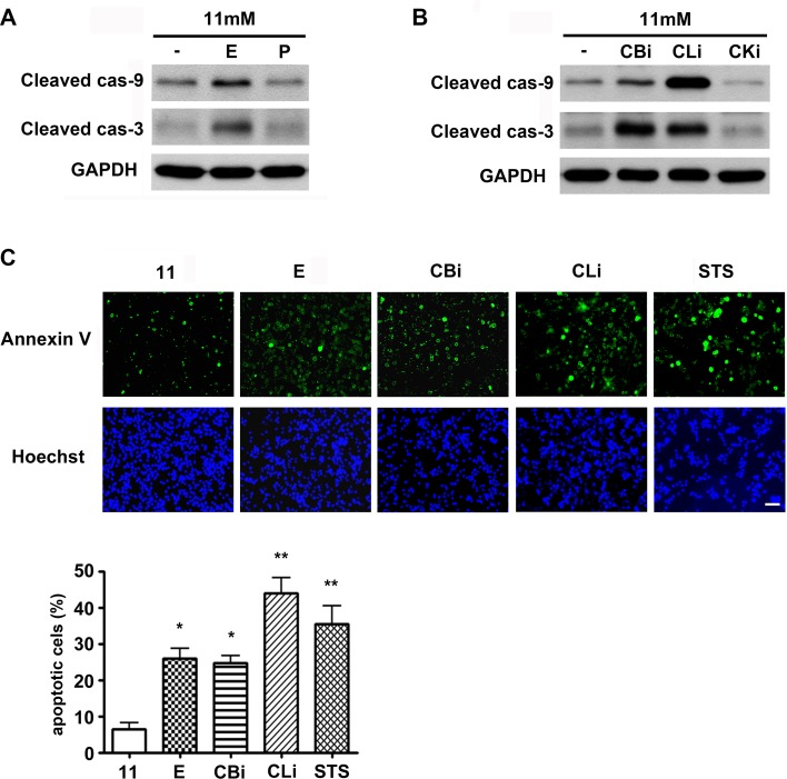
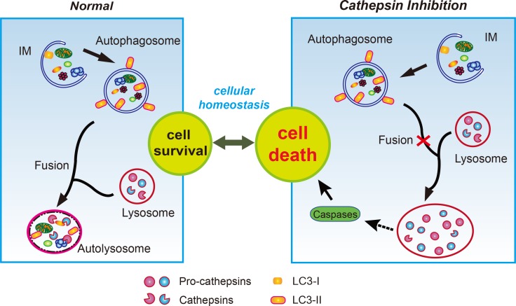
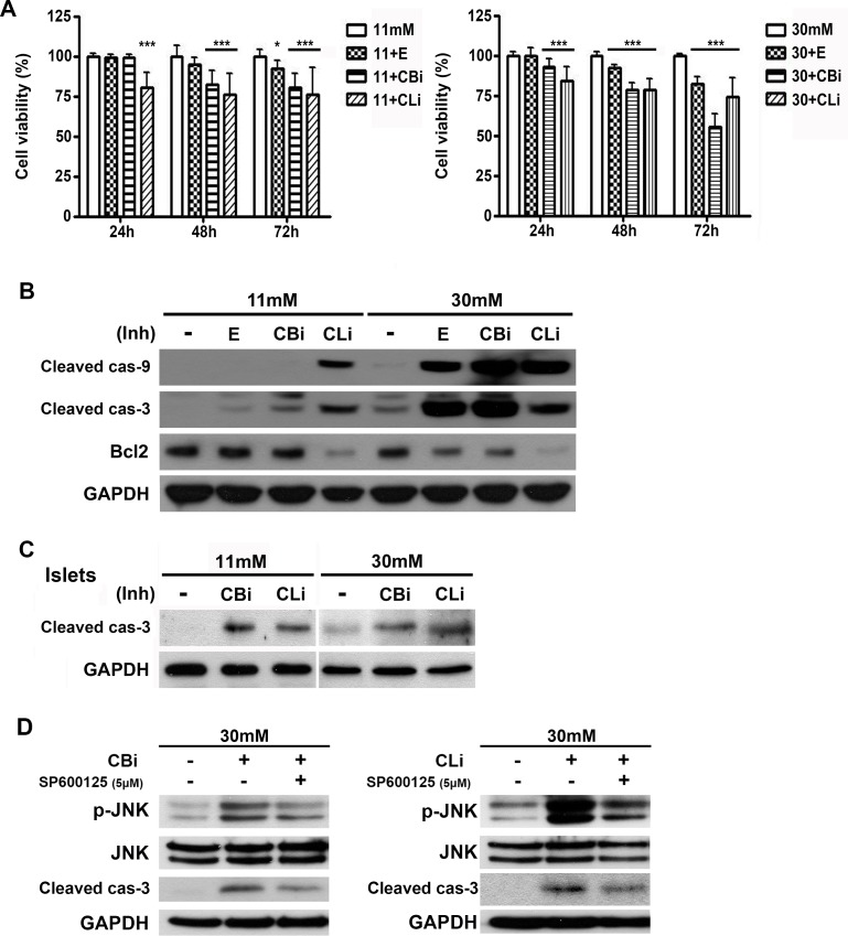
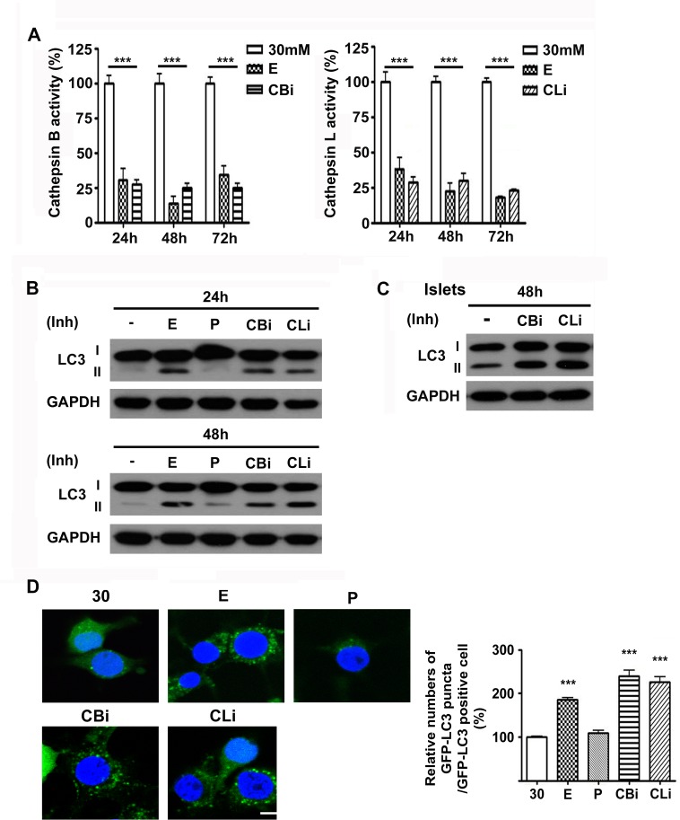
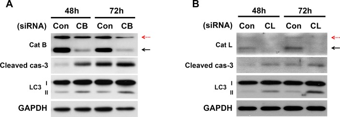
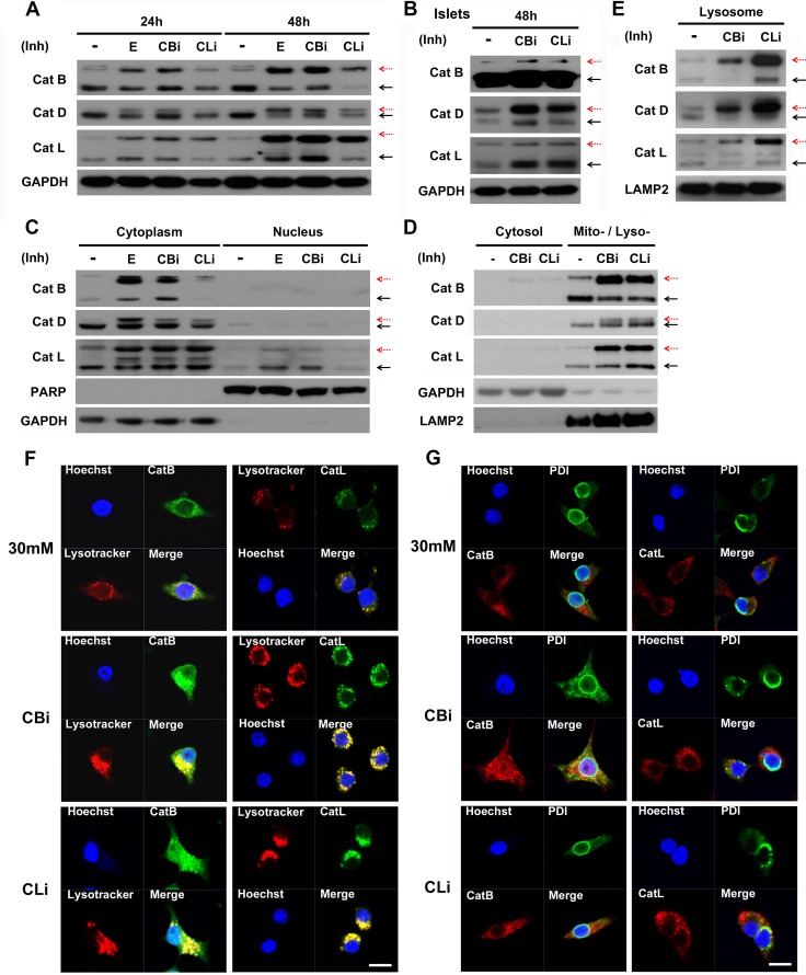
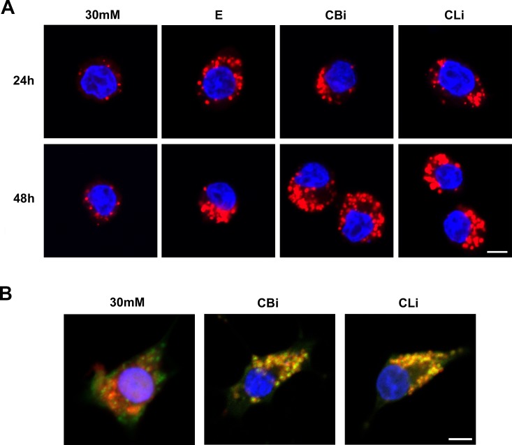
Autophagy is a lysosomal degradative pathway that plays an important role in maintaining cellular homeostasis. We previously showed that the inhibition of autophagy causes pancreatic β-cell apoptosis, suggesting that autophagy is a protective mechanism for the survival of pancreatic β-cells. The current study demonstrates that treatment with inhibitors and knockdown of the lysosomal cysteine proteases such as cathepsins B and L impair autophagy, enhancing the caspase-dependent apoptosis of INS-1 cells and islets upon exposure to high concentration of glucose. Interestingly, treatment with cathepsin B and L inhibitors prevented the proteolytic processing of cathepsins B, D and L, as evidenced by gradual accumulation of the respective pro-forms. Of note, inhibition of aspartic cathepsins had no effect on autophagy and cell viability, suggesting the selective role of cathepsins B and L in the regulation of β-cell autophagy and apoptosis. Lysosomal localization of accumulated pro-cathepsins in the presence of cathepsin B and L inhibitors was verified via immunocytochemistry and lysosomal fractionation. Lysotracker staining indicated that cathepsin B and L inhibitors led to the formation of severely enlarged lysosomes in a time-dependent manner. The abnormal accumulation of pro-cathepsins following treatment with inhibitors of cathepsins B and L suppressed normal lysosomal degradation and the processing of lysosomal enzymes, leading to lysosomal dysfunction. Collectively, our findings suggest that cathepsin defects following the inhibition of cathepsin B and L result in lysosomal dysfunction and consequent cell death in pancreatic β-cells.
自噬是一种溶酶体降解途径,在维持细胞内稳态中发挥重要作用。我们之前的研究表明,抑制自噬会导致胰腺β细胞凋亡,这表明自噬是胰腺β细胞存活的一种保护机制。当前研究表明,用抑制剂处理以及敲低组织蛋白酶B和L等溶酶体半胱氨酸蛋白酶会损害自噬,增强INS-1细胞和胰岛在高浓度葡萄糖作用下依赖半胱天冬酶的凋亡。有趣的是,用组织蛋白酶B和L抑制剂处理可阻止组织蛋白酶B、D和L的蛋白水解加工,各自前体形式的逐渐积累证明了这一点。值得注意的是,抑制天冬氨酸组织蛋白酶对自噬和细胞活力没有影响,这表明组织蛋白酶B和L在β细胞自噬和凋亡调节中具有选择性作用。通过免疫细胞化学和溶酶体分级分离证实了在存在组织蛋白酶B和L抑制剂的情况下积累的前体组织蛋白酶的溶酶体定位。溶酶体示踪剂染色表明,组织蛋白酶B和L抑制剂以时间依赖性方式导致形成严重增大的溶酶体。用组织蛋白酶B和L抑制剂处理后前体组织蛋白酶的异常积累抑制了正常的溶酶体降解和溶酶体酶的加工,导致溶酶体功能障碍。总的来说,我们的研究结果表明,抑制组织蛋白酶B和L后组织蛋白酶缺陷会导致胰腺β细胞溶酶体功能障碍并随之发生细胞死亡。