McDonald A J, Zaric V
Department of Pharmacology, Physiology and Neuroscience, University of South Carolina School of Medicine, Columbia, SC 29208, United States.
Department of Pharmacology, Physiology and Neuroscience, University of South Carolina School of Medicine, Columbia, SC 29208, United States.
Neuroscience. 2015 Apr 2;290:227-42. doi: 10.1016/j.neuroscience.2015.01.028. Epub 2015 Jan 28.
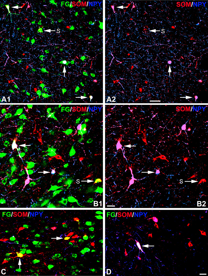
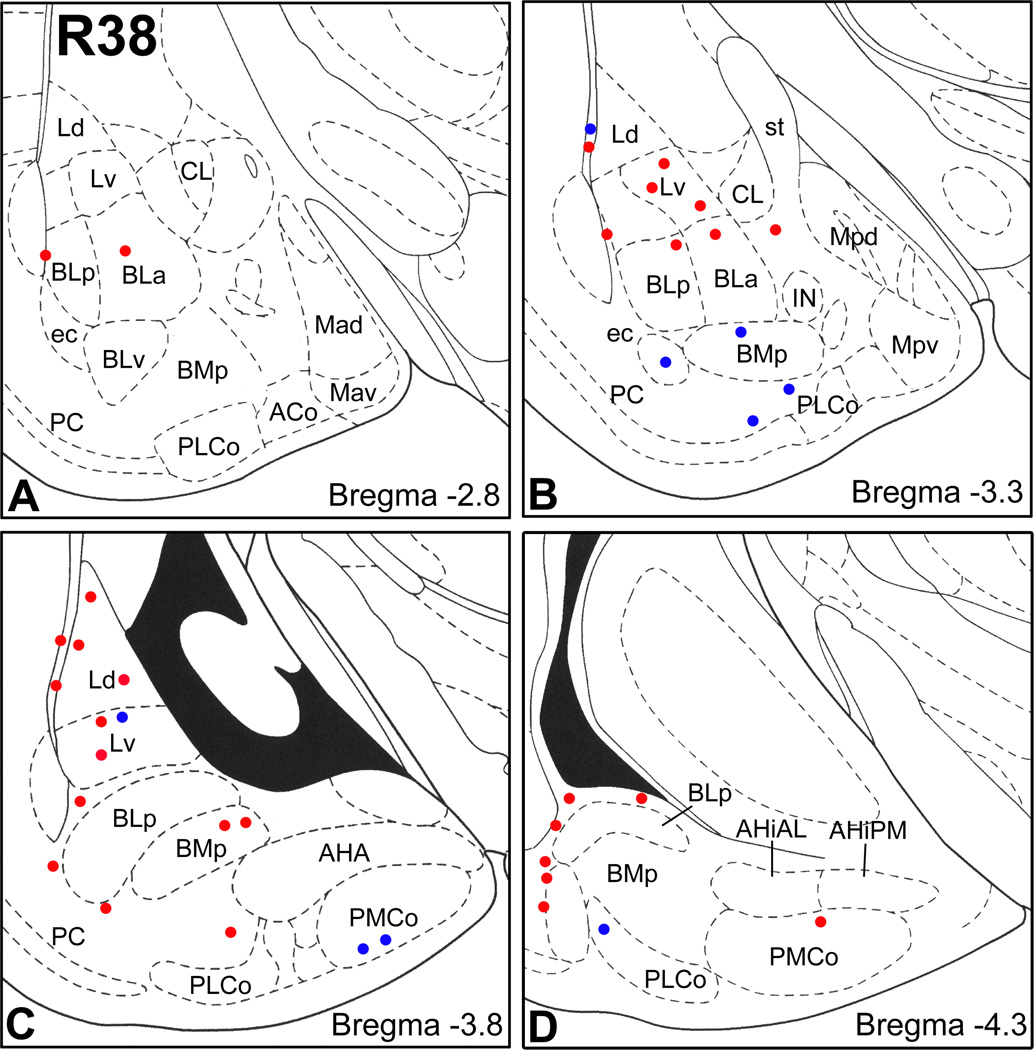
The entorhinal cortex and other hippocampal and parahippocampal cortices are interconnected by a small number of GABAergic nonpyramidal neurons in addition to glutamatergic pyramidal cells. Since the cortical and basolateral amygdalar nuclei have cortex-like cell types and have robust projections to the entorhinal cortex, we hypothesized that a small number of amygdalar GABAergic nonpyramidal neurons might participate in amygdalo-entorhinal projections. To test this hypothesis we combined Fluorogold (FG) retrograde tract tracing with immunohistochemistry for the amygdalar nonpyramidal cell markers glutamic acid decarboxylase (GAD), parvalbumin (PV), somatostatin (SOM), neuropeptide Y (NPY), vasoactive intestinal peptide (VIP), and the m2 muscarinic cholinergic receptor (M2R). Injections of FG into the rat entorhinal cortex labeled numerous neurons that were mainly located in the cortical and basolateral nuclei of the amygdala. Although most of these amygdalar FG+ neurons labeled by entorhinal injections were large pyramidal cells, 1-5% were smaller long-range nonpyramidal neurons (LRNP neurons) that expressed SOM, or both SOM and NPY. No amygdalar FG+ neurons in these cases were PV+ or VIP+. Cell counts revealed that LRNP neurons labeled by injections into the entorhinal cortex constituted about 10-20% of the total SOM+ population, and 20-40% of the total NPY population in portions of the lateral amygdalar nucleus that exhibited a high density of FG+ neurons. Sixty-two percent of amygdalar FG+/SOM+ neurons were GAD+, and 51% were M2R+. Since GABAergic projection neurons typically have low perikaryal levels of GABAergic markers, it is actually possible that most or all of the amygdalar LRNP neurons are GABAergic. Like GABAergic LRNP neurons in hippocampal/parahippocampal regions, amygdalar LRNP neurons that project to the entorhinal cortex are most likely involved in synchronizing oscillatory activity between the two regions. These oscillations could entrain synchronous firing of amygdalar and entorhinal pyramidal neurons, thus facilitating functional interactions between them, including synaptic plasticity.
除了谷氨酸能锥体细胞外,内嗅皮质与其他海马及海马旁回皮质通过少数γ-氨基丁酸能(GABAergic)非锥体神经元相互连接。由于皮质杏仁核和基底外侧杏仁核具有类似皮质的细胞类型,并且对内嗅皮质有强大的投射,我们推测少数杏仁核GABA能非锥体神经元可能参与杏仁核-内嗅投射。为了验证这一假设,我们将荧光金(FG)逆行束路追踪与免疫组织化学相结合,检测杏仁核非锥体细胞标志物谷氨酸脱羧酶(GAD)、小白蛋白(PV)、生长抑素(SOM)、神经肽Y(NPY)、血管活性肠肽(VIP)和M2型毒蕈碱胆碱能受体(M2R)。向大鼠内嗅皮质注射FG标记了大量神经元,这些神经元主要位于杏仁核的皮质核和基底外侧核。虽然内嗅注射标记的这些杏仁核FG+神经元大多数是大型锥体细胞,但1-5%是较小的长程非锥体神经元(LRNP神经元),它们表达SOM,或同时表达SOM和NPY。在这些情况下,没有杏仁核FG+神经元是PV+或VIP+。细胞计数显示被注射到内嗅皮质标记的LRNP神经元在外侧杏仁核中FG+神经元高密度区域约占SOM+细胞总数的10-20%,占NPY细胞总数的20-40%。62%的杏仁核FG+/SOM+神经元是GAD+,51%是M2R+。由于GABA能投射神经元通常在胞体中GABA能标志物水平较低,实际上大多数或所有杏仁核LRNP神经元都可能是GABA能的。与海马/海马旁回区域的GABA能LRNP神经元一样,投射到内嗅皮质的杏仁核LRNP神经元很可能参与两个区域之间振荡活动的同步。这些振荡可能带动杏仁核和内嗅锥体细胞的同步放电,从而促进它们之间的功能相互作用,包括突触可塑性。