Acta Neuropathol Commun. 2015 Feb 3;3:9. doi: 10.1186/s40478-015-0189-z.
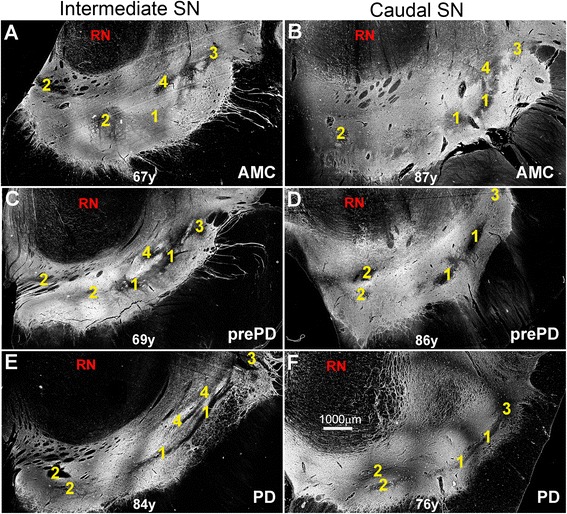
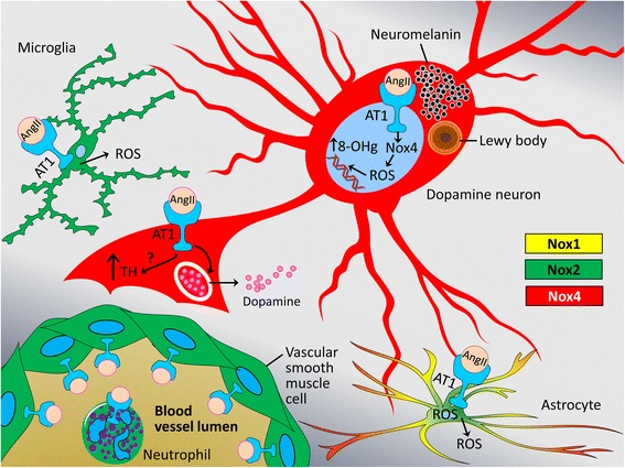
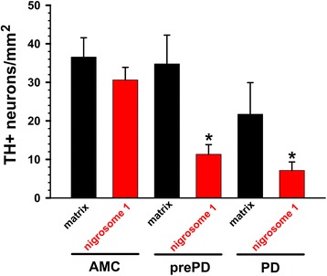
In rodent models of Parkinson's disease (PD), dopamine neuron loss is accompanied by increased expression of angiotensin II (AngII), its type 1 receptor (AT1), and NADPH oxidase (Nox) in the nigral dopamine neurons and microglia. AT1 blockers (ARBs) stymie such oxidative damage and neuron loss. Whether changes in the AngII/AT1/Nox4 axis contribute to Parkinson neuropathogenesis is unknown. Here, we studied the distribution of AT1 and Nox4 in dopamine neurons in two nigral subregions: the less affected calbindin-rich matrix and the first-affected calbindin-poor nigrosome 1 of three patients, who were clinically asymptomatic, but had nigral dopamine cell loss and Braak stages consistent with a neuropathological diagnosis of PD (prePD). For comparison, five clinically- and neuropathologically-confirmed PD patients and seven age-matched control patients (AMC) were examined.
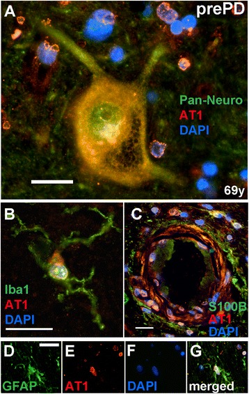
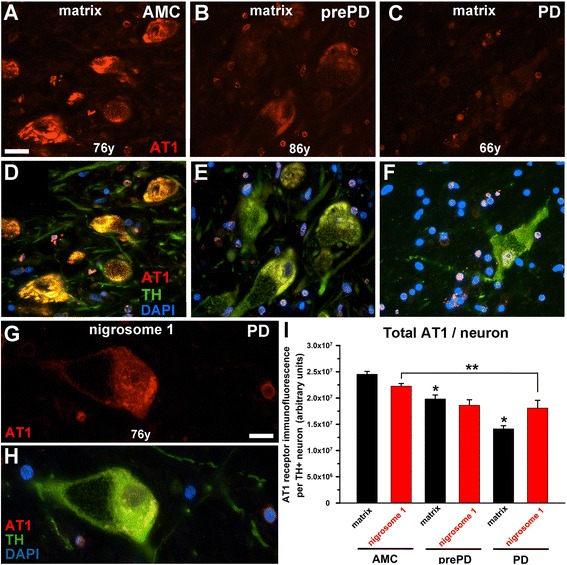
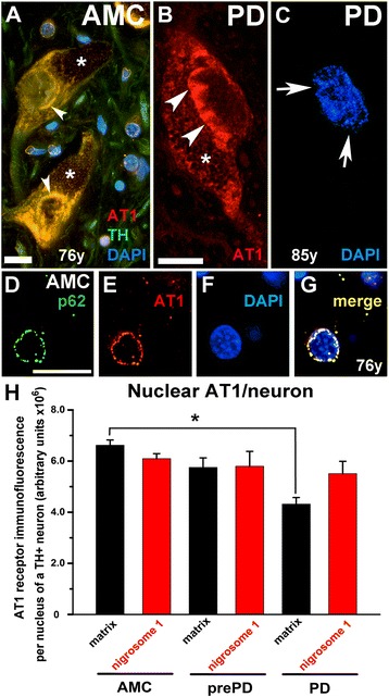
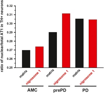
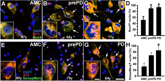
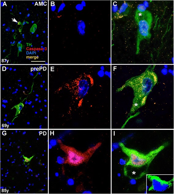
AT1 and Nox4 immunoreactivity was noted in dopamine neurons in both the matrix and the nigrosome 1. The total cellular levels of AT1 in surviving dopamine neurons in the matrix and nigrosome 1 declined from AMC>prePD>PD, suggesting that an AngII/AT1/Nox4 axis orders neurodegenerative progression. In this vein, the loss of dopamine neurons was paralleled by a decline in total AT1 per surviving dopamine neuron. Similarly, AT1 in the nuclei of surviving neurons in the nigral matrix declined with disease progression, i.e., AMC>prePD>PD. In contrast, in nigrosome 1, the expression of nuclear AT1 was unaffected and similar in all groups. The ratio of nuclear AT1 to total AT1 (nuclear + cytoplasmic + membrane) in dopamine neurons increased stepwise from AMC to prePD to PD. The proportional increase in nuclear AT1 in dopamine neurons in nigrosome 1 of prePD and PD patients was accompanied by elevated nuclear expression of Nox4, oxidative damage to DNA, and caspase-3-mediated cell loss.
Our observations are consistent with the idea that AngII/AT1/Nox4 axis-mediated oxidative stress gives rise to the dopamine neuron dysfunction and loss characteristic of the neuropathological and clinical manifestations of PD and suggest that the chance for a neuron to survive increases in association with lower total as well as nuclear AT1 expression. Our results support the need for further evaluation of ARBs as disease-modifying agents in PD.
在帕金森病(PD)的啮齿动物模型中,血管紧张素 II(AngII)、其 1 型受体(AT1)和 NADPH 氧化酶(Nox)在黑质多巴胺神经元和小胶质细胞中的表达增加,伴随着多巴胺神经元的丢失。AT1 阻滞剂(ARB)抑制了这种氧化损伤和神经元丢失。AngII/AT1/Nox4 轴的变化是否导致帕金森神经病变尚不清楚。在这里,我们研究了 AT1 和 Nox4 在两个黑质亚区多巴胺神经元中的分布:三个临床无症状但黑质多巴胺细胞丢失和 Braak 阶段与帕金森病的病理诊断一致(prePD)的患者。为了比较,检查了五名临床和病理确诊的 PD 患者和七名年龄匹配的对照患者(AMC)。
AT1 和 Nox4 免疫反应性在基质和 nigrosome 1 中的多巴胺神经元中均有发现。基质和 nigrosome 1 中存活的多巴胺神经元中总细胞 AT1 水平从 AMC>prePD>PD 下降,提示 AngII/AT1/Nox4 轴控制神经退行性进展。在这方面,多巴胺神经元的丢失伴随着每个存活的多巴胺神经元中总 AT1 的减少。同样,黑质基质中存活神经元的核 AT1 随疾病进展而下降,即 AMC>prePD>PD。相比之下,nigrosome 1 中的核 AT1 表达不受影响,在所有组中都相似。从 AMC 到 prePD 再到 PD,多巴胺神经元中核 AT1 与总 AT1(核+细胞质+膜)的比值逐步增加。prePD 和 PD 患者 nigrosome 1 中多巴胺神经元核 AT1 的比例增加伴随着 Nox4 的核表达升高、DNA 氧化损伤和 caspase-3 介导的细胞丢失。
我们的观察结果与 AngII/AT1/Nox4 轴介导的氧化应激导致多巴胺神经元功能障碍和丢失的观点一致,这种功能障碍和丢失是 PD 的神经病理学和临床表现的特征,并表明神经元存活的机会随着总 AT1 和核 AT1 表达的降低而增加。我们的结果支持进一步评估 ARB 作为 PD 的疾病修饰治疗的需要。