Histomorphometry and Stereology Research Centre, Shiraz University of Medical Sciences, Shiraz, Iran. ; Anatomy Department, School of Medicine, Shiraz University of Medical Sciences, Shiraz, Iran.
Anatomy Department, School of Medicine, Shiraz University of Medical Sciences, Shiraz, Iran.
Psychiatry Investig. 2015 Jan;12(1):73-80. doi: 10.4306/pi.2015.12.1.73. Epub 2015 Jan 12.
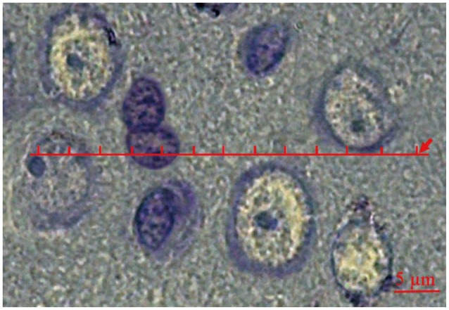
The present study explored the three-dimensional spatial arrangements of the neurons and glial cells within the medial prefrontal cortex (mPFC) of rats.
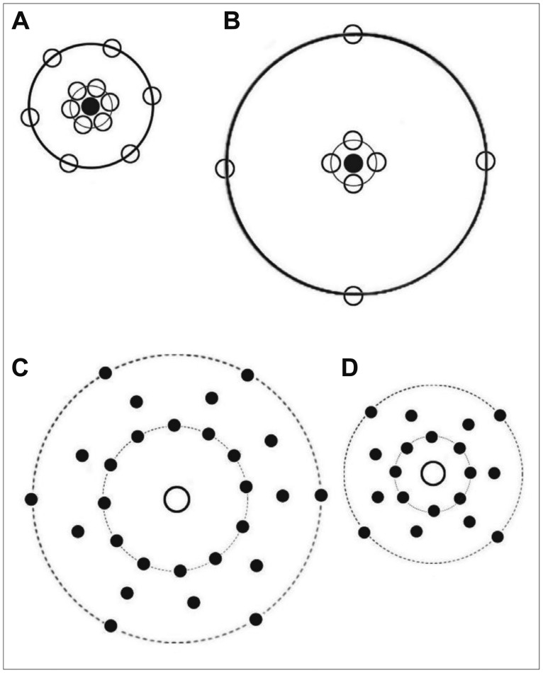
It evaluated the arrangement for differences after stress with or without treatment with curcumin and sertraline using second-order stereology. Orientator method was applied to obtain isotropic uniform random sections of mPFC. The pair correlation g(r) and cross-correlation functions were estimated by counting dipole probes superimposed on histological sections of mPFC.
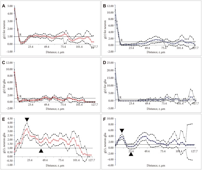
The mean total volume of neurons and glial cells was 0.80 (0.05) and 0.40 (0.07), respectively in the control group. The corresponding values decreased by 50% in the stressed group. The curve of g(r) for the neurons and glial cells showed a wider gap between the stressed rats' mPFC. Theses indicate a negative correlation (repulsion) between the neurons and glial cells in the stressed rats. Evaluation of the cross-correlation function of the neurons and glial cells also showed a negative correlation in the stressed group. The estimated values of the global degree of order in the spatial point pattern for neurons and glial cells were 0.62 and 0.20 in control and stressed animals, respectively. Curcumin and sertraline protected the spatial arrangements of the cells after stress induction in rats. In addition, the volume of the neurons and glial cells remained unchanged after stress.
Dissociation of the neurons and glial cells can is seen at some places in the stressed rats' cortex. However, the spatial arrangement of the cells was remained unchanged in curcumin+stress and sertraline+stress rats.
本研究探索了大鼠前额皮质内侧区(mPFC)神经元和神经胶质细胞的三维空间排列。
采用二级体视学法评估应激后给予姜黄素和舍曲林治疗的大鼠mPFC 排列差异。应用定向方法获得 mPFC 的各向同性均匀随机切片。通过在 mPFC 的组织切片上叠加偶极探针来估计对相关函数 g(r)和交叉相关函数。
对照组神经元和神经胶质细胞的平均总体积分别为 0.80(0.05)和 0.40(0.07)。应激组相应值下降 50%。神经元和神经胶质细胞的 g(r)曲线显示应激大鼠 mPFC 之间的间隙更大。这表明应激大鼠的神经元和神经胶质细胞之间存在负相关(排斥)。神经元和神经胶质细胞的交叉相关函数评估也显示应激组存在负相关。神经元和神经胶质细胞的空间点模式全局有序度的估计值分别为 0.62 和 0.20 在对照组和应激组动物中。姜黄素和舍曲林在应激诱导后保护了大鼠细胞的空间排列。此外,应激后神经元和神经胶质细胞的体积保持不变。
应激大鼠皮质的某些部位可见神经元和神经胶质细胞的分离。然而,姜黄素+应激和舍曲林+应激大鼠的细胞空间排列保持不变。