Budell Lesley, Kunz Miriam, Jackson Philip L, Rainville Pierre
Département de physiologie, Université de Montréal, Montréal, Québec, Canada; Groupe de recherche sur le système nerveux central (GRSNC) and Centre de recherche de l'Institut universitaire de gériatrie de Montréal (CRIUGM), Montréal, Québec, Canada.
Department of Psychology, University of Bamberg, Bamberg, Germany.
PLoS One. 2015 Feb 11;10(2):e0107526. doi: 10.1371/journal.pone.0107526. eCollection 2015.
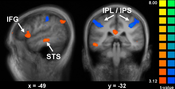
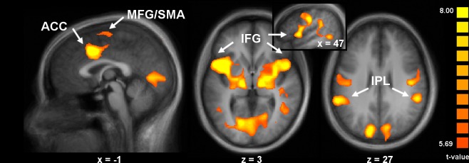
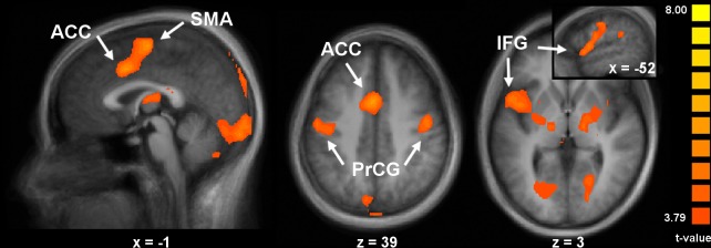
Perception of pain in others via facial expressions has been shown to involve brain areas responsive to self-pain, biological motion, as well as both performed and observed motor actions. Here, we investigated the involvement of these different regions during emotional and motor mirroring of pain expressions using a two-task paradigm, and including both observation and execution of the expressions. BOLD responses were measured as subjects watched video clips showing different intensities of pain expression and, after a variable delay, either expressed the amount of pain they perceived in the clips (pain task), or imitated the facial movements (movement task). In the pain task condition, pain coding involved overlapping activation across observation and execution in the anterior cingulate cortex, supplementary motor area, inferior frontal gyrus/anterior insula, and the inferior parietal lobule, and a pain-related increase (pain vs. neutral) in the anterior cingulate cortex/supplementary motor area, the right inferior frontal gyrus, and the postcentral gyrus. The 'mirroring' response was stronger in the inferior frontal gyrus and middle temporal gyrus/superior temporal sulcus during the pain task, and stronger in the inferior parietal lobule in the movement task. These results strongly suggest that while motor mirroring may contribute to the perception of pain expressions in others, interpreting these expressions in terms of pain content draws more heavily on networks involved in the perception of affective meaning.
通过面部表情感知他人的疼痛已被证明涉及对自身疼痛、生物运动以及执行和观察到的运动动作有反应的脑区。在这里,我们使用双任务范式,研究了在疼痛表情的情感和运动镜像过程中这些不同区域的参与情况,包括对表情的观察和执行。当受试者观看显示不同强度疼痛表情的视频片段时,测量其血氧水平依赖(BOLD)反应,并在可变延迟后,受试者要么表达他们在片段中感知到的疼痛程度(疼痛任务),要么模仿面部动作(运动任务)。在疼痛任务条件下,疼痛编码涉及前扣带回皮质、辅助运动区、额下回/前脑岛和顶下小叶在观察和执行过程中的重叠激活,以及前扣带回皮质/辅助运动区、右侧额下回和中央后回中与疼痛相关的增加(疼痛与中性相比)。在疼痛任务期间,额下回以及颞中回/颞上沟的“镜像”反应更强,而在运动任务中,顶下小叶的“镜像”反应更强。这些结果强烈表明,虽然运动镜像可能有助于对他人疼痛表情的感知,但根据疼痛内容来解释这些表情更多地依赖于参与情感意义感知的网络。