Department of Biomedical Engineering, University of Utah, Salt Lake City, Utah, United States of America.
Department of Neurosurgery, University of Utah, Salt Lake City, Utah, United States of America.
PLoS One. 2023 Oct 16;18(10):e0292808. doi: 10.1371/journal.pone.0292808. eCollection 2023.
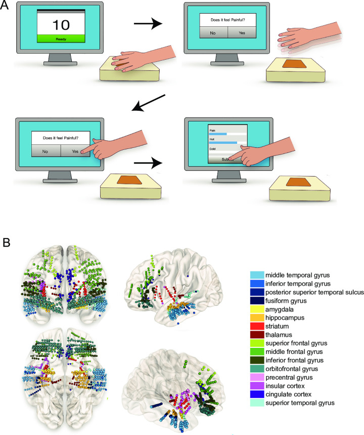
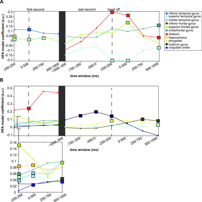
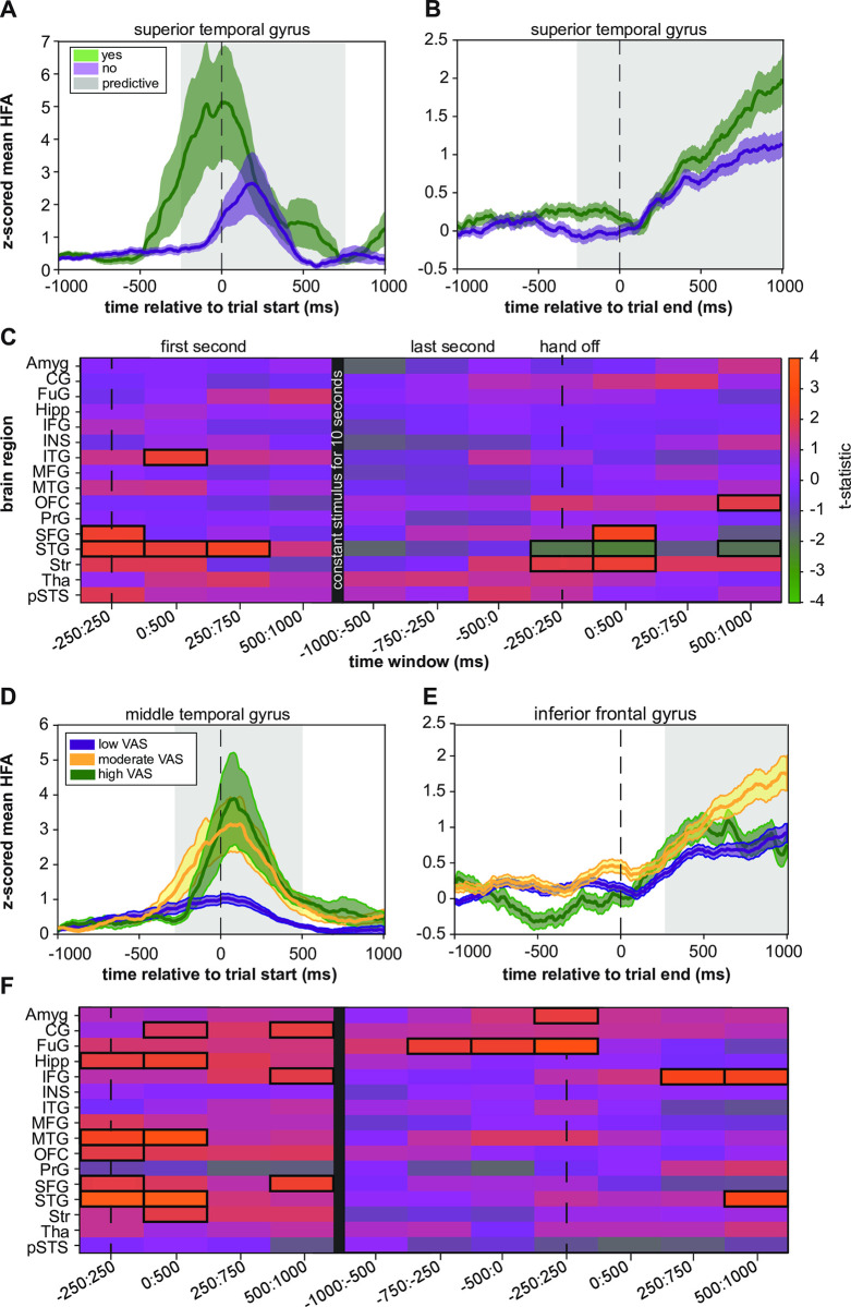
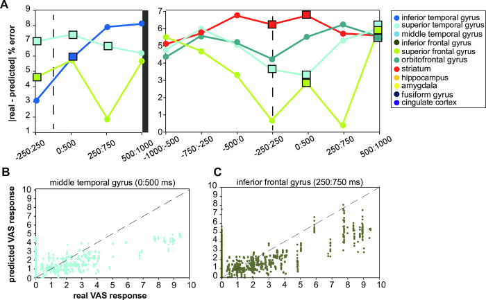
Pain is a complex experience involving sensory, emotional, and cognitive aspects, and multiple networks manage its processing in the brain. Examining how pain transforms into a behavioral response can shed light on the networks' relationships and facilitate interventions to treat chronic pain. However, studies using high spatial and temporal resolution methods to investigate the neural encoding of pain and its psychophysical correlates have been limited. We recorded from intracranial stereo-EEG (sEEG) electrodes implanted in sixteen different brain regions of twenty patients who underwent psychophysical pain testing consisting of a tonic thermal stimulus to the hand. Broadband high-frequency local field potential amplitude (HFA; 70-150 Hz) was isolated to investigate the relationship between the ongoing neural activity and the resulting psychophysical pain evaluations. Two different generalized linear mixed-effects models (GLME) were employed to assess the neural representations underlying binary and graded pain psychophysics. The first model examined the relationship between HFA and whether the patient responded "yes" or "no" to whether the trial was painful. The second model investigated the relationship between HFA and how painful the stimulus was rated on a visual analog scale. GLMEs revealed that HFA in the inferior temporal gyrus (ITG), superior frontal gyrus (SFG), and superior temporal gyrus (STG) predicted painful responses at stimulus onset. An increase in HFA in the orbitofrontal cortex (OFC), SFG, and striatum predicted pain responses at stimulus offset. Numerous regions, including the anterior cingulate cortex, hippocampus, IFG, MTG, OFC, and striatum, predicted the pain rating at stimulus onset. However, only the amygdala and fusiform gyrus predicted increased pain ratings at stimulus offset. We characterized the spatiotemporal representations of binary and graded painful responses during tonic pain stimuli. Our study provides evidence from intracranial recordings that the neural encoding of psychophysical pain changes over time during a tonic thermal stimulus, with different brain regions being predictive of pain at the beginning and end of the stimulus.
疼痛是一种复杂的体验,涉及感觉、情感和认知方面,多个网络在大脑中管理其处理。研究疼痛如何转化为行为反应可以揭示网络之间的关系,并有助于干预慢性疼痛。然而,使用高空间和时间分辨率方法研究疼痛的神经编码及其心理物理学相关性的研究一直受到限制。我们从接受包括手部持续热刺激的心理物理学疼痛测试的二十名患者的十六个不同大脑区域植入的颅内立体脑电图 (sEEG) 电极中进行了记录。我们分离了宽带高频局部场电势幅度 (HFA;70-150 Hz),以研究持续神经活动与由此产生的心理物理学疼痛评估之间的关系。我们采用了两种不同的广义线性混合效应模型 (GLME) 来评估二进制和分级疼痛心理物理学的神经表示。第一个模型检查了 HFA 与患者对试验是否疼痛的“是”或“否”反应之间的关系。第二个模型研究了 HFA 与视觉模拟量表上对刺激的疼痛评分之间的关系。GLME 显示,颞下回 (ITG)、额上回 (SFG) 和颞上回 (STG) 的 HFA 预测了刺激开始时的疼痛反应。眶额皮层 (OFC)、SFG 和纹状体的 HFA 增加预测了刺激结束时的疼痛反应。包括前扣带皮层、海马体、IFG、MTG、OFC 和纹状体在内的许多区域都预测了刺激开始时的疼痛评分。然而,只有杏仁核和梭状回预测了刺激结束时疼痛评分的增加。我们描述了在持续热刺激期间二进制和分级疼痛反应的时空表示。我们的研究提供了颅内记录的证据,表明在持续热刺激期间,心理物理学疼痛的神经编码随时间而变化,不同的大脑区域可预测刺激开始和结束时的疼痛。