• 文献检索
  • 文档翻译
  • 深度研究
  • 学术资讯
  • Suppr Zotero 插件Zotero 插件
  • 邀请有礼
  • 套餐&价格
  • 历史记录
应用&插件
Suppr Zotero 插件Zotero 插件浏览器插件Mac 客户端Windows 客户端微信小程序
定价
高级版会员购买积分包购买API积分包
服务
文献检索文档翻译深度研究API 文档MCP 服务
关于我们
关于 Suppr公司介绍联系我们用户协议隐私条款
关注我们

Suppr 超能文献

核心技术专利:CN118964589B侵权必究
粤ICP备2023148730 号-1Suppr @ 2026

文献检索

告别复杂PubMed语法,用中文像聊天一样搜索,搜遍4000万医学文献。AI智能推荐,让科研检索更轻松。

立即免费搜索

文件翻译

保留排版,准确专业,支持PDF/Word/PPT等文件格式,支持 12+语言互译。

免费翻译文档

深度研究

AI帮你快速写综述,25分钟生成高质量综述,智能提取关键信息,辅助科研写作。

立即免费体验

中美儿童及青少年大脑发育轨迹的比较。

Comparison of the brain development trajectory between Chinese and U.S. children and adolescents.

作者信息

Xie Wanze, Richards John E, Lei Du, Lee Kang, Gong Qiyong

机构信息

Department of Psychology, Institute for Mind and Brain, University of South Carolina Columbia, SC, USA ; Huaxi MR Research Center (HMRRC), Department of Radiology, West China Hospital of Sichuan University Chengdu, China.

Department of Psychology, Institute for Mind and Brain, University of South Carolina Columbia, SC, USA.

出版信息

Front Syst Neurosci. 2015 Feb 2;8:249. doi: 10.3389/fnsys.2014.00249. eCollection 2014.

DOI:10.3389/fnsys.2014.00249
PMID:25698941
原文链接:https://pmc.ncbi.nlm.nih.gov/articles/PMC4313711/
Abstract

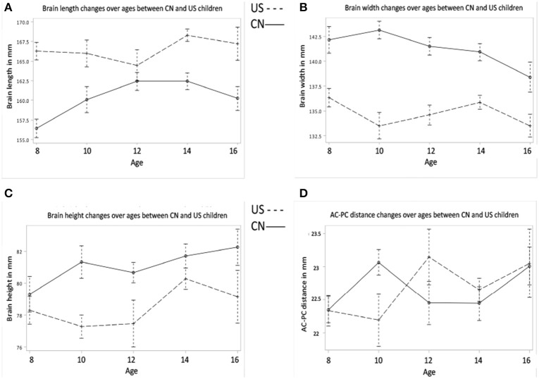
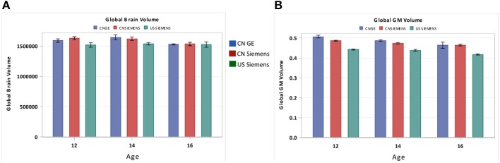
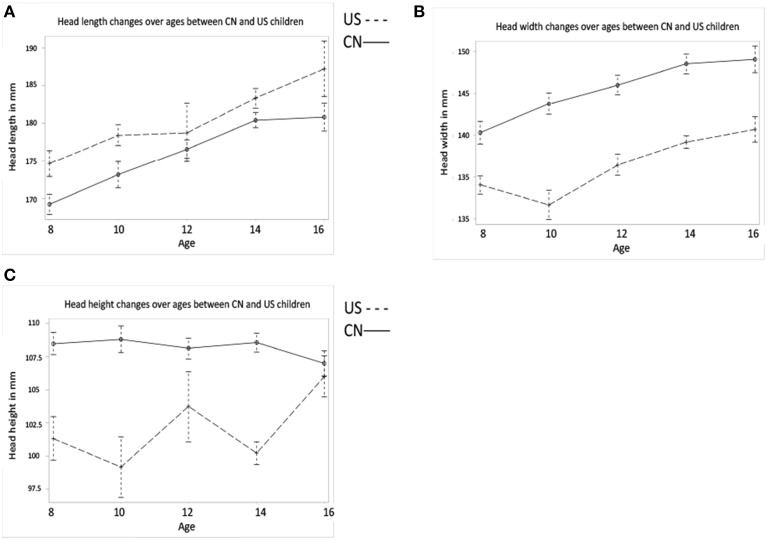
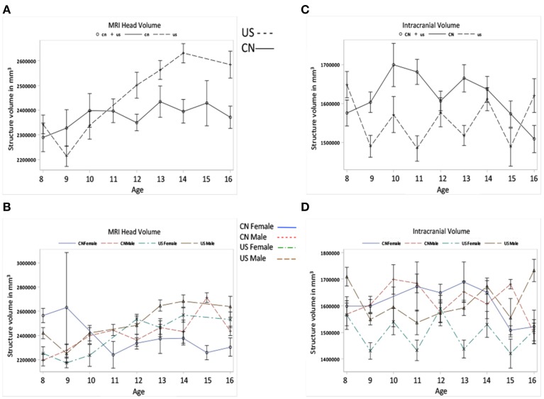
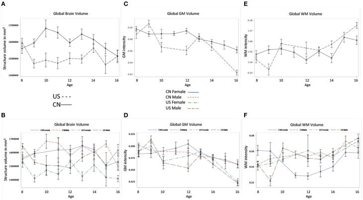
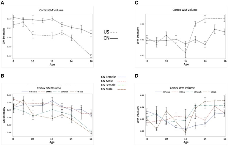
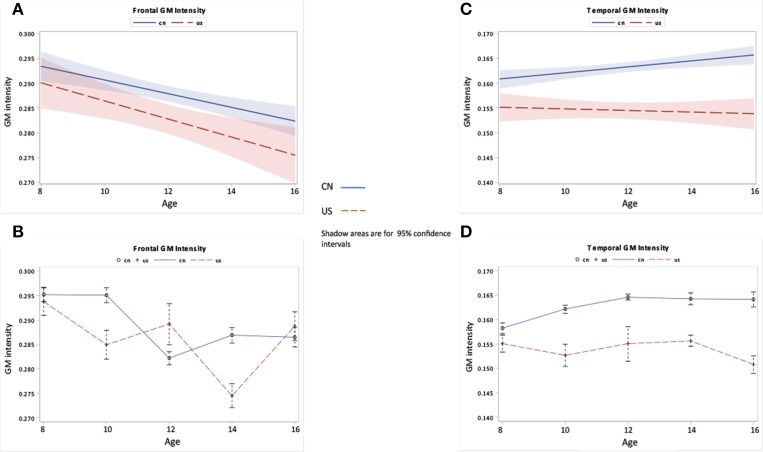
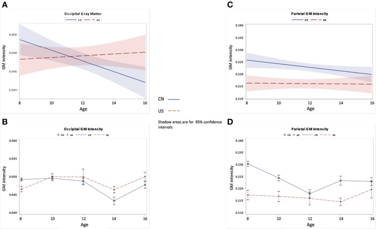
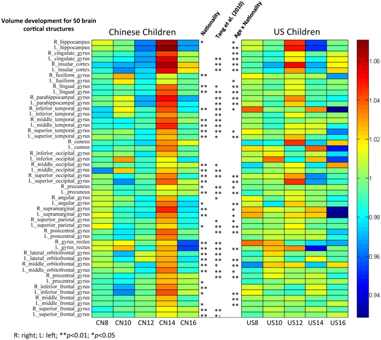
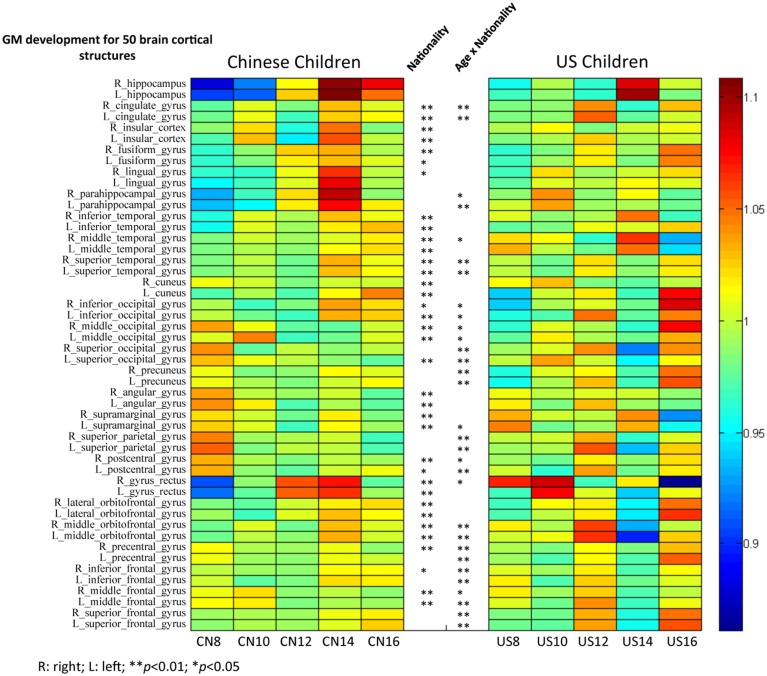
This current study investigated brain development of Chinese and American children and adolescents from 8 to 16 years of age using structural magnetic resonance imaging (MRI) techniques. Analyses comparing Chinese and U.S. children brain/head MR images were performed to explore similarities and differences in the trajectory of brain development between these two groups. Our results revealed regional and age differences in both brain/head morphological and tissue level development between Chinese and U.S. children. Chinese children's brains and heads were shorter, wider, and taller than those of U.S. children. There were significant differences in the gray matter (GM) and white matter (WM) intensity between the two nationalities. Development trajectories for cerebral volume, GM, and several key brain structures were also distinct between these two populations.

摘要

本研究采用结构磁共振成像(MRI)技术,对8至16岁的中国和美国儿童及青少年的大脑发育情况进行了调查。通过分析比较中国和美国儿童的脑/头部磁共振图像,探索这两组人群大脑发育轨迹的异同。我们的研究结果显示,中国和美国儿童在脑/头部形态和组织水平发育方面存在区域和年龄差异。中国儿童的大脑和头部比美国儿童的更短、更宽、更高。两国儿童在灰质(GM)和白质(WM)强度上存在显著差异。这两个人群在脑容量、GM和几个关键脑结构的发育轨迹上也有所不同。

https://cdn.ncbi.nlm.nih.gov/pmc/blobs/3782/4313711/d9624d2038c3/fnsys-08-00249-g0010.jpg
https://cdn.ncbi.nlm.nih.gov/pmc/blobs/3782/4313711/1e84530606e6/fnsys-08-00249-g0001.jpg
https://cdn.ncbi.nlm.nih.gov/pmc/blobs/3782/4313711/67d8cc43f499/fnsys-08-00249-g0002.jpg
https://cdn.ncbi.nlm.nih.gov/pmc/blobs/3782/4313711/76fae6b82a1a/fnsys-08-00249-g0003.jpg
https://cdn.ncbi.nlm.nih.gov/pmc/blobs/3782/4313711/30e3cc184785/fnsys-08-00249-g0004.jpg
https://cdn.ncbi.nlm.nih.gov/pmc/blobs/3782/4313711/581aac362797/fnsys-08-00249-g0005.jpg
https://cdn.ncbi.nlm.nih.gov/pmc/blobs/3782/4313711/8c7dbea5c728/fnsys-08-00249-g0006.jpg
https://cdn.ncbi.nlm.nih.gov/pmc/blobs/3782/4313711/6b60e599e125/fnsys-08-00249-g0007.jpg
https://cdn.ncbi.nlm.nih.gov/pmc/blobs/3782/4313711/63cb9dafaa34/fnsys-08-00249-g0008.jpg
https://cdn.ncbi.nlm.nih.gov/pmc/blobs/3782/4313711/2d8061380a34/fnsys-08-00249-g0009.jpg
https://cdn.ncbi.nlm.nih.gov/pmc/blobs/3782/4313711/d9624d2038c3/fnsys-08-00249-g0010.jpg

相似文献

1
Comparison of the brain development trajectory between Chinese and U.S. children and adolescents.中美儿童及青少年大脑发育轨迹的比较。
Front Syst Neurosci. 2015 Feb 2;8:249. doi: 10.3389/fnsys.2014.00249. eCollection 2014.
2
The construction of MRI brain/head templates for Chinese children from 7 to 16 years of age.7至16岁中国儿童的脑部磁共振成像(MRI)模板构建。
Dev Cogn Neurosci. 2015 Oct;15:94-105. doi: 10.1016/j.dcn.2015.08.008. Epub 2015 Aug 28.
3
Structural brain differences in school-age children with and without single-suture craniosynostosis.患有和未患有单缝颅缝早闭症的学龄儿童的大脑结构差异。
J Neurosurg Pediatr. 2017 Apr;19(4):479-489. doi: 10.3171/2016.9.PEDS16107. Epub 2017 Feb 3.
4
Brain Magnetic Resonance Imaging Findings in Children and Young Adults With CKD.儿童和青少年慢性肾脏病患者的脑部磁共振成像结果。
Am J Kidney Dis. 2018 Sep;72(3):349-359. doi: 10.1053/j.ajkd.2017.11.024. Epub 2018 Feb 15.
5
Brain development in Chinese children and adolescents: a structural MRI study.中国儿童和青少年的脑发育:一项结构磁共振成像研究。
Neuroreport. 2007 Jun 11;18(9):875-80. doi: 10.1097/WNR.0b013e328152777e.
6
A reliable spatially normalized template of the human spinal cord--Applications to automated white matter/gray matter segmentation and tensor-based morphometry (TBM) mapping of gray matter alterations occurring with age.一个可靠的人类脊髓空间标准化模板——应用于自动白质/灰质分割以及基于张量的形态测量学(TBM)对与年龄相关的灰质变化进行映射。
Neuroimage. 2015 Aug 15;117:20-8. doi: 10.1016/j.neuroimage.2015.05.034. Epub 2015 May 21.
7
White-matter lesions drive deep gray-matter atrophy in early multiple sclerosis: support from structural MRI.白质病变驱动早期多发性硬化症的深部灰质萎缩:来自结构 MRI 的支持。
Mult Scler. 2013 Oct;19(11):1485-92. doi: 10.1177/1352458513478673. Epub 2013 Mar 5.
8
Comparison of whole brain segmentation and volume estimation in children and young adults using SPM and SyMRI.SPM 和 SyMRI 比较儿童和青年大脑整体分割和体积估计。
Clin Imaging. 2019 Sep-Oct;57:77-82. doi: 10.1016/j.clinimag.2019.05.008. Epub 2019 May 21.
9
Characterization of gray-matter multiple sclerosis lesions using double inversion recovery, diffusion, contrast-enhanced, and volumetric MRI.采用双反转恢复、弥散、对比增强和容积 MRI 对灰质多发性硬化病变进行特征描述。
Mult Scler Relat Disord. 2019 Jun;31:74-81. doi: 10.1016/j.msard.2019.03.021. Epub 2019 Mar 29.
10
Adiposity is associated with structural properties of the adolescent brain.肥胖与青少年大脑的结构特性有关。
Neuroimage. 2014 Dec;103:192-201. doi: 10.1016/j.neuroimage.2014.09.030. Epub 2014 Sep 26.

引用本文的文献

1
Population-specific brain charts reveal Chinese-Western differences in neurodevelopmental trajectories.特定人群脑图谱揭示了中西神经发育轨迹的差异。
bioRxiv. 2025 Jun 18:2025.06.17.659820. doi: 10.1101/2025.06.17.659820.
2
A three-step, "brute-force" approach toward optimized affine spatial normalization.一种用于优化仿射空间归一化的三步“强力”方法。
Front Comput Neurosci. 2024 Jul 8;18:1367148. doi: 10.3389/fncom.2024.1367148. eCollection 2024.
3
Specific-CT brain template construction and retrospective dosimetric comparison study in brain for nasopharyngeal carcinoma patients treated with IMRT or VMAT.

本文引用的文献

1
The autism brain imaging data exchange: towards a large-scale evaluation of the intrinsic brain architecture in autism.自闭症脑影像数据交换:走向自闭症内在大脑结构的大规模评估。
Mol Psychiatry. 2014 Jun;19(6):659-67. doi: 10.1038/mp.2013.78. Epub 2013 Jun 18.
2
Age-specific MRI templates for pediatric neuroimaging.用于儿科神经影像学的特定年龄MRI模板。
Dev Neuropsychol. 2012;37(5):379-99. doi: 10.1080/87565641.2012.688900.
3
FSL.束流输送系统。
鼻咽癌患者接受调强放疗(IMRT)或容积旋转调强放疗(VMAT)治疗时脑部特异性CT模板构建及回顾性剂量学比较研究
Am J Cancer Res. 2024 Apr 15;14(4):1662-1674. doi: 10.62347/HACN9549. eCollection 2024.
4
A detailed dosimetric comparative study of IMRT and VMAT in normal brain tissues for nasopharyngeal carcinoma patients treated with radiotherapy.鼻咽癌患者放疗中调强放疗(IMRT)与容积调强弧形放疗(VMAT)在正常脑组织中的详细剂量学对比研究。
Front Radiol. 2023 May 18;3:1190763. doi: 10.3389/fradi.2023.1190763. eCollection 2023.
5
A Chinese multi-modal neuroimaging data release for increasing diversity of human brain mapping.中国人脑多模态神经影像数据发布增加人脑图谱研究的多样性。
Sci Data. 2022 Jun 9;9(1):286. doi: 10.1038/s41597-022-01413-3.
6
Cortical thinning and sleep slow wave activity reductions mediate age-related improvements in cognition during mid-late adolescence.皮质变薄和睡眠慢波活动减少介导了从中年到青春期后期认知能力随年龄增长而提高。
Sleep. 2022 Jan 11;45(1). doi: 10.1093/sleep/zsab206.
7
Little evidence for associations between the Big Five personality traits and variability in brain gray or white matter.大五人格特质与大脑灰质或白质变异性之间的关联证据不足。
Neuroimage. 2020 Oct 15;220:117092. doi: 10.1016/j.neuroimage.2020.117092. Epub 2020 Jun 27.
8
Constructing and evaluating a cortical surface atlas and analyzing cortical sex differences in young Chinese adults.构建和评估中国人皮质表面图谱及分析中国青年大脑皮质性别差异
Hum Brain Mapp. 2020 Jun 15;41(9):2495-2513. doi: 10.1002/hbm.24960. Epub 2020 Mar 6.
9
Sample sizes and population differences in brain template construction.脑模板构建中的样本量和人群差异。
Neuroimage. 2020 Feb 1;206:116318. doi: 10.1016/j.neuroimage.2019.116318. Epub 2019 Nov 2.
10
Localizing Age-Related Changes in Brain Structure Using Voxel-Based Morphometry.使用基于体素的形态测量法定位大脑结构中与年龄相关的变化。
Neural Plast. 2017;2017:6303512. doi: 10.1155/2017/6303512. Epub 2017 Jan 17.
Neuroimage. 2012 Aug 15;62(2):782-90. doi: 10.1016/j.neuroimage.2011.09.015. Epub 2011 Sep 16.
4
Neurodevelopmental MRI brain templates for children from 2 weeks to 4 years of age.神经发育磁共振成像脑模板,适用于 2 周至 4 岁的儿童。
Dev Psychobiol. 2012 Jan;54(1):77-91. doi: 10.1002/dev.20579. Epub 2011 Jun 17.
5
A comparison between Chinese and Caucasian head shapes.中国人与高加索人头型的比较。
Appl Ergon. 2010 Oct;41(6):832-9. doi: 10.1016/j.apergo.2010.02.002. Epub 2010 Mar 12.
6
The construction of a Chinese MRI brain atlas: a morphometric comparison study between Chinese and Caucasian cohorts.中国人脑 MRI 图谱构建:中国人与高加索人群队列的形态计量学比较研究。
Neuroimage. 2010 May 15;51(1):33-41. doi: 10.1016/j.neuroimage.2010.01.111. Epub 2010 Feb 10.
7
Bayesian analysis of neuroimaging data in FSL.基于FSL的神经影像数据的贝叶斯分析。
Neuroimage. 2009 Mar;45(1 Suppl):S173-86. doi: 10.1016/j.neuroimage.2008.10.055. Epub 2008 Nov 13.
8
Automatic segmentation of brain MRIs of 2-year-olds into 83 regions of interest.将2岁幼儿的脑部磁共振成像自动分割为83个感兴趣区域。
Neuroimage. 2008 Apr 1;40(2):672-684. doi: 10.1016/j.neuroimage.2007.11.034. Epub 2007 Dec 3.
9
Construction of a 3D probabilistic atlas of human cortical structures.人类皮质结构三维概率图谱的构建。
Neuroimage. 2008 Feb 1;39(3):1064-80. doi: 10.1016/j.neuroimage.2007.09.031. Epub 2007 Nov 26.
10
Brain development in Chinese children and adolescents: a structural MRI study.中国儿童和青少年的脑发育:一项结构磁共振成像研究。
Neuroreport. 2007 Jun 11;18(9):875-80. doi: 10.1097/WNR.0b013e328152777e.