Research Center for Sectional and Imaging Anatomy, Shandong University School of Medicine, Jinan, Shandong 250012, China.
Neuroimage. 2010 May 15;51(1):33-41. doi: 10.1016/j.neuroimage.2010.01.111. Epub 2010 Feb 10.
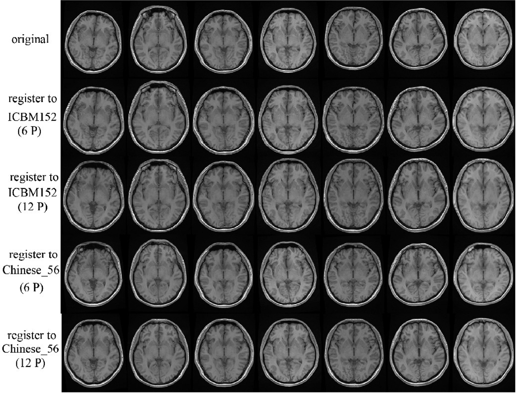
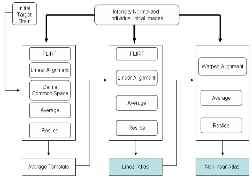
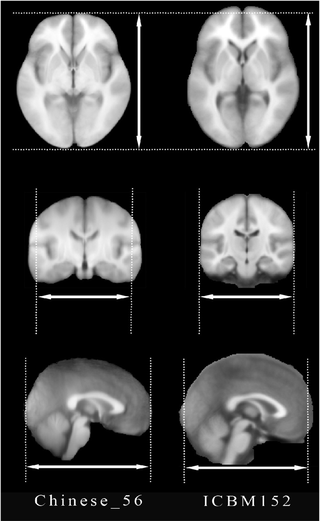
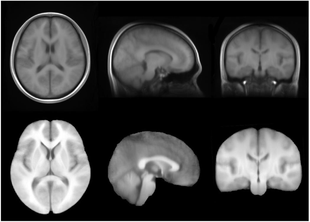
We developed a novel brain atlas template to facilitate computational brain studies of Chinese subjects and populations using high quality magnetic resonance imaging (MRI) and well-validated image analysis techniques. To explore the ethnicity-based structural brain differences, we used the MRI scans of 35 Chinese male subjects (24.03+/-2.06 years) and compared them to an age-matched cohort of 35 Caucasian males (24.03+/-2.06 years). Global volumetric measures were used to identify significant group differences in the brain length, width, height and AC-PC line distance. Using the LONI BrainParser, 56 brain structures were automatically labeled and analyzed for all subjects. We identified significant ethnicity differences in brain structure volumes, suggesting that a population-specific brain atlas may be more appropriate for studies involving Chinese populations. To address this, we constructed a 3D Chinese brain atlas based on high resolution 3.0T MRI scans of 56 right-handed male Chinese volunteers (24.46+/-1.81 years). All Chinese brains were spatially normalized by using linear and nonlinear transformation via the "AIR Make Atlas" pipeline workflow within the LONI pipeline environment. This high-resolution Chinese brain atlas was compared to the ICBM152 template, which was constructed using Caucasian brains.
我们开发了一种新的大脑图谱模板,以使用高质量磁共振成像(MRI)和经过良好验证的图像分析技术,促进中国受试者和人群的计算性大脑研究。为了探索基于种族的结构脑差异,我们使用了 35 名中国男性受试者(24.03+/-2.06 岁)的 MRI 扫描,并将其与年龄匹配的 35 名白种男性(24.03+/-2.06 岁)进行了比较。使用全局体积测量来确定大脑长度、宽度、高度和 AC-PC 线距离的显著组间差异。使用 LONI BrainParser,自动标记和分析所有受试者的 56 个大脑结构。我们发现大脑结构体积存在显著的种族差异,这表明特定于人群的大脑图谱可能更适合涉及中国人群的研究。为了解决这个问题,我们根据 56 名右利手中国男性志愿者(24.46+/-1.81 岁)的高分辨率 3.0T MRI 扫描构建了一个 3D 中国大脑图谱。通过 LONI 管道环境中的“AIR Make Atlas”管道工作流程,使用线性和非线性变换对所有中国大脑进行空间归一化。这个高分辨率的中国大脑图谱与使用白种人大脑构建的 ICBM152 模板进行了比较。