Ok Evren, Adanir Necdet, Ozturk Tuba
Department of Endodontics, Faculty of Dentistry, Sifa University, Izmir, Turkiye.
Department of Endodontics, Faculty of Dentistry, Suleyman Demirel University, Isparta, Turkiye.
Eur J Dent. 2015 Jan-Mar;9(1):20-24. doi: 10.4103/1305-7456.149633.
The aim of this in vitro study was to evaluate the antimicrobial effect of oregano extract solution (OES) against Enterococcus faecalis within root canals and dentin tubules, and its effect on smear layer.
A total of 180 human maxillary central incisors was selected. After removal of coronal part of the teeth, root canals were prepared using ProTaper rotary files (Dentsply, Tulsa Endodontics, OK, USA) to #F3 with the crown-down manner. The roots were randomly assigned to 15 groups (n = 12 for each). In the first seven groups, the antimicrobial effects of the test groups were evaluated. Suspensions of E. faecalis cultures were adjusted to 1.0 McFarland (1 × 10(8) colony-forming unit [CFU]/ml), and sterilized teeth were placed in Eppendorf tubes and kept at 37°C for 4 weeks. Samples were then taken from the root canals before irrigation using three sterile paper points. Dentin samples were taken from root canals with ProTaper #F4 and #F5 series rotary instruments after irrigation. The aliquots of samples were placed into the brain heart infusion and incubated at 37°C for 48 h and then the CFUs were counted. In the other eight groups, the efficacy of the irrigation solutions on removing the smear layer was evaluated using scanning electron microscope (Leo 440, Oxford Microscopy Ltd., Cambridge, England) analysis. Statistical evaluation of the microbiological data was performed using the Kruskall-Wallis and Mann-Witney U-test (P < 0.05).
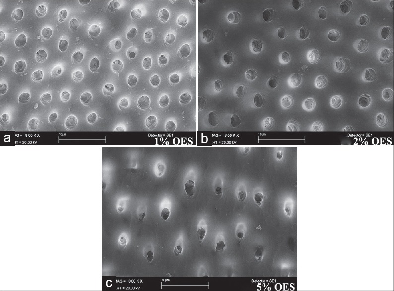
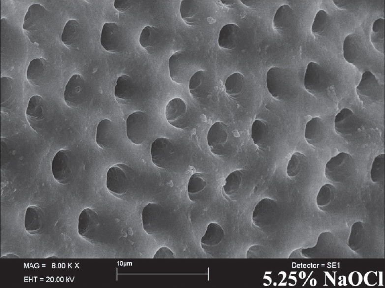
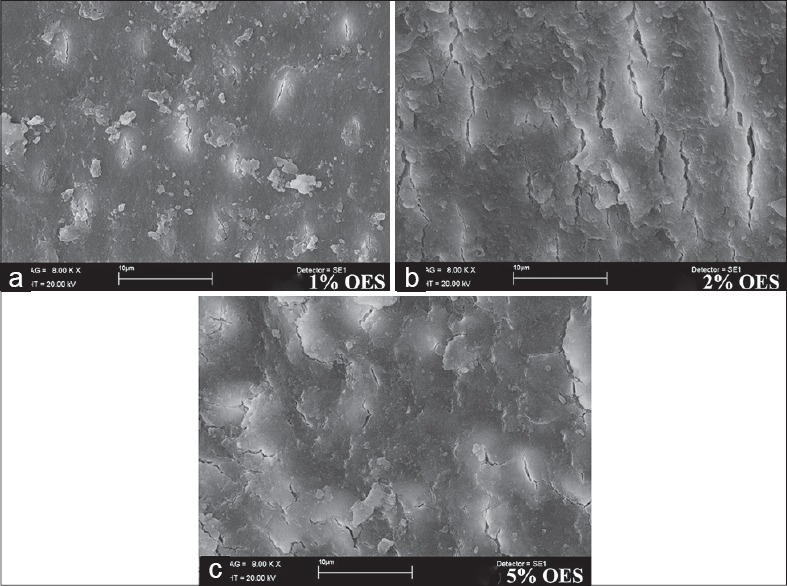
There was a statistically difference between the groups (P < 0.05). Chlorhexidine gluconate (CHX), 5% and 2% OES wasn't found to be statistically significant regarding their antibacterial activities against E. faecalis (P > 0.05). 1% OES and NaOCl showed similar antimicrobial effect (P > 0.05), and 1% OES and NaOCl were better than ethylenediaminetetraacetic acid (EDTA) and saline (P < 0.05) but not as successful as CHX. According to the results obtained from dentin, CHX is the most effective solution within dentinal tubules. Different concentrations of OES were not achieved smear layer removal alone but OES in conjunction with 17% EDTA was the final irrigating solution achieved the smear layer removal without dentin erosion.
Within the limitations of this study, OES appears to be a possible alternative to NaOCl as a root canal irrigant on the eradication of E. faecalis and removal of smear layer.
本体外研究旨在评估牛至提取物溶液(OES)对根管和牙本质小管内粪肠球菌的抗菌作用及其对玷污层的影响。
共选取180颗人上颌中切牙。去除牙冠部分后,使用ProTaper旋转锉(美国登士柏公司,塔尔萨牙髓病学公司,俄克拉何马州)以冠向下法将根管预备至F3号。将牙根随机分为15组(每组n = 12)。在前七组中,评估试验组的抗菌效果。将粪肠球菌培养物悬液调整至1.0麦氏浊度(1×10⁸菌落形成单位[CFU]/ml),将消毒后的牙齿置于Eppendorf管中,在37°C下保存4周。然后在冲洗前使用三个无菌纸尖从根管中取样。冲洗后,使用ProTaper F4和F5系列旋转器械从根管中获取牙本质样本。将样本等分试样置于脑心浸液中,在37°C下孵育48小时,然后计数CFU。在其他八组中,使用扫描电子显微镜(英国剑桥牛津显微镜有限公司的Leo 440)分析评估冲洗液去除玷污层的效果。使用Kruskal-Wallis和Mann-Whitney U检验对微生物学数据进行统计学评估(P < 0.05)。
各组之间存在统计学差异(P < 0.05)。葡萄糖酸氯己定(CHX)、5%和2%的OES对粪肠球菌的抗菌活性未发现有统计学意义(P > 0.05)。1%的OES和次氯酸钠(NaOCl)显示出相似的抗菌效果(P > 0.05),1%的OES和NaOCl优于乙二胺四乙酸(EDTA)和生理盐水(P < 0.05),但不如CHX有效。根据从牙本质获得的结果,CHX是牙本质小管内最有效的溶液。不同浓度的OES单独不能去除玷污层,但OES与17%的EDTA联合作为最终冲洗液可去除玷污层且不会造成牙本质侵蚀。
在本研究的局限性内,OES似乎可作为NaOCl的一种可能替代物,用于根管冲洗以根除粪肠球菌和去除玷污层。