Xie Jia-Xin, Feng Yu, Yuan Jian-Min, You Zhen-Dong, Lin Hai-Yan, Lu Chang-Lin, Xu Jia-Jun
Department of Anatomy, The Second Military Medical University, Shanghai, P. R. China; Department of Orthopaedics, Changzheng Hospital, The Second Military Medical University, Shanghai, P. R. China; People's Liberation Army Clinical Center for Spinal Cord Injury, Kunming General Hospital of Chengdu Military Command, Kunming, P. R. China.
People's Liberation Army Clinical Center for Spinal Cord Injury, Kunming General Hospital of Chengdu Military Command, Kunming, P. R. China.
PLoS One. 2015 Mar 3;10(3):e0119119. doi: 10.1371/journal.pone.0119119. eCollection 2015.
Effective therapy for visual loss caused by optic nerve injury or diseases has not been achieved even though the optic nerve has the regeneration potential after injury. This study was designed to modify amniotic epithelial cells (AECs) with basic fibroblast growth factor (bFGF) gene, preliminarily investigating its effect on transected optic nerve.
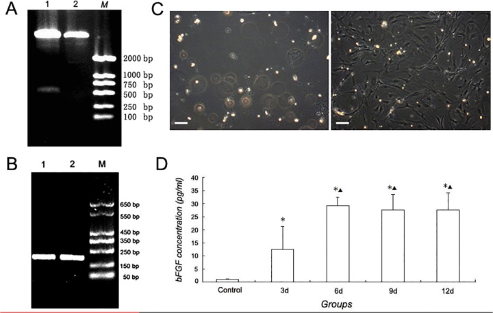
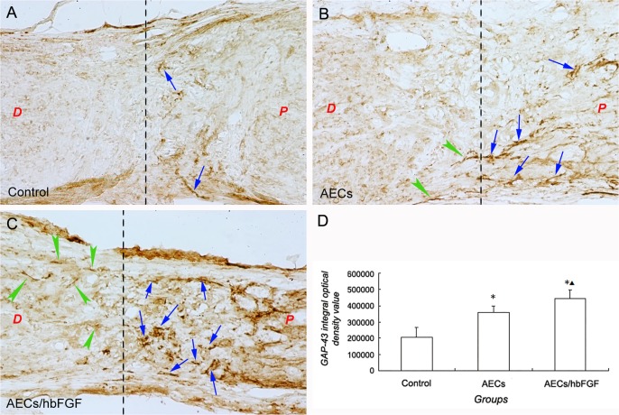
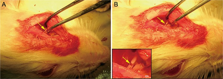
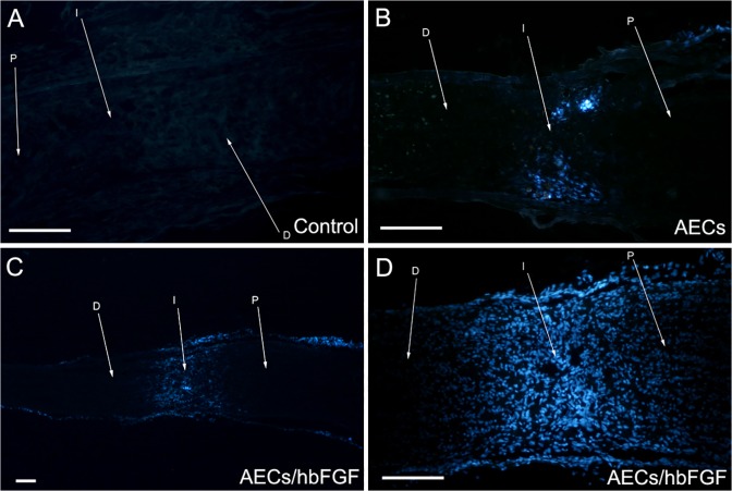
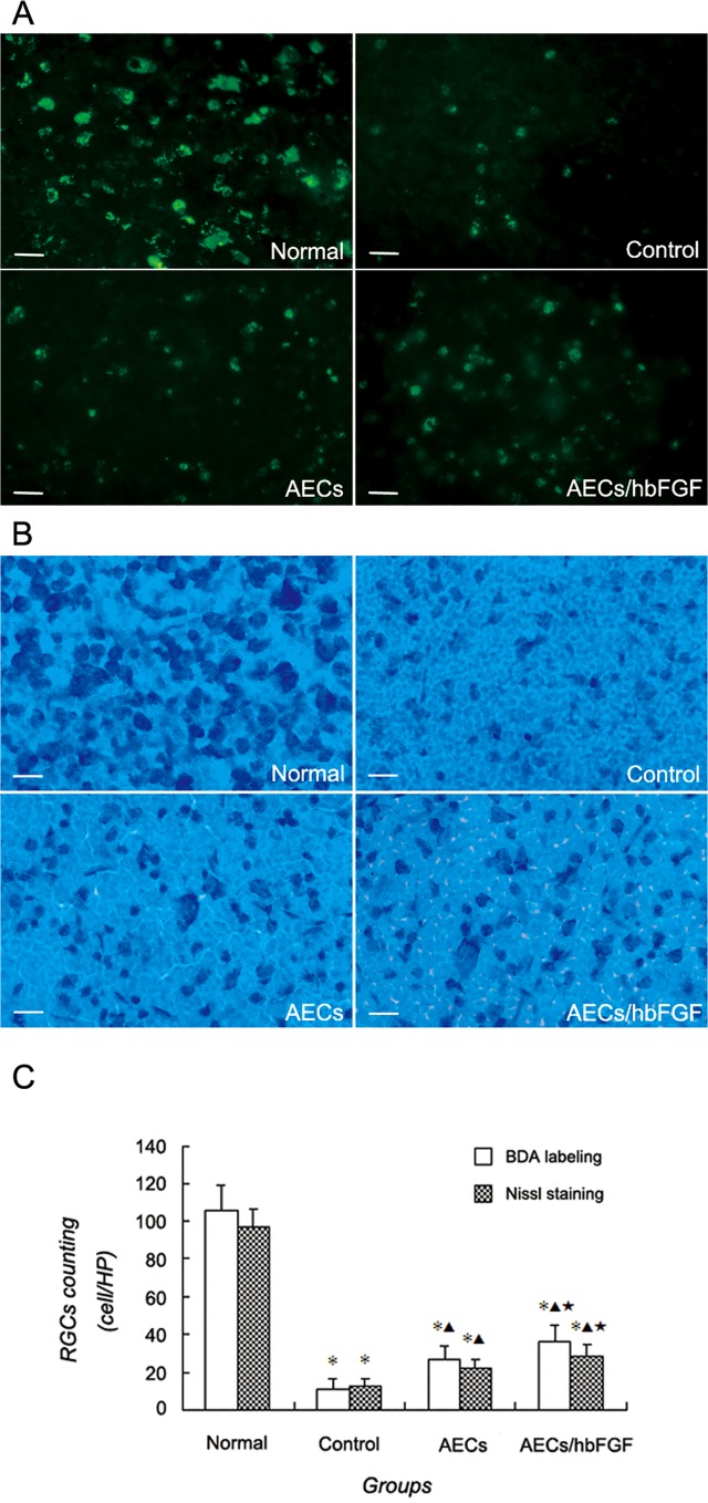
A human bFGF gene segment was delivered into rat AECs (AECs/hbFGF) by lentiviral vector, and the gene expression was examined by RT-PCR and ELISA. The AECs/hbFGF and untransfected rat AECs were transplanted into the transected site of the rat optic nerve. At 28 days post transplantation, the survival and migration of the transplanted cells was observed by tracking labeled cells; meanwhile retinal ganglion cells (RGCs) were observed and counted by employing biotin dextran amine (BDA) and Nissl staining. Furthermore, the expression of growth associated protein 43 (GAP-43) within the injury site was examined with immunohistochemical staining.
The AECs/hbFGF was proven to express bFGF gene and secrete bFGF peptide. Both AECs/hbFGF and AECs could survive and migrate after transplantation. RGCs counting implicated that RGCs numbers of the cell transplantation groups were significantly higher than that of the control group, and the AECs/hbFGF group was significantly higher than that of the AECs group. Moreover GAP-43 integral optical density value in the control group was significantly lower than that of the cell transplantation groups, and the value in the AECs/hbFGF group was significantly higher than that of the AECs group.
AECs modified with bFGF could reduce RGCs loss and promote expression of GAP-43 in the rat optic nerve transected model, facilitating the process of neural restoration following injury.
尽管视神经损伤后具有再生潜力,但尚未实现对视神经损伤或疾病所致视力丧失的有效治疗。本研究旨在用碱性成纤维细胞生长因子(bFGF)基因修饰羊膜上皮细胞(AECs),初步研究其对横断视神经的作用。
通过慢病毒载体将人bFGF基因片段导入大鼠AECs(AECs/hbFGF),采用逆转录聚合酶链反应(RT-PCR)和酶联免疫吸附测定(ELISA)检测基因表达。将AECs/hbFGF和未转染的大鼠AECs移植到大鼠视神经横断部位。移植后28天,通过追踪标记细胞观察移植细胞的存活和迁移情况;同时,采用生物素葡聚糖胺(BDA)和尼氏染色法观察并计数视网膜神经节细胞(RGCs)。此外,用免疫组织化学染色法检测损伤部位生长相关蛋白43(GAP-43)的表达。
证实AECs/hbFGF表达bFGF基因并分泌bFGF肽。AECs/hbFGF和AECs移植后均能存活并迁移。RGCs计数表明,细胞移植组的RGCs数量显著高于对照组,且AECs/hbFGF组显著高于AECs组。此外,对照组GAP-43积分光密度值显著低于细胞移植组,AECs/hbFGF组的值显著高于AECs组。
用bFGF修饰的AECs可减少大鼠视神经横断模型中RGCs的损失,并促进GAP-43在损伤部位的表达,从而促进损伤后神经修复进程。