Kostova Elena B, Beuger Boukje M, Klei Thomas R L, Halonen Pasi, Lieftink Cor, Beijersbergen Roderick, van den Berg Timo K, van Bruggen Robin
*Department of Blood Cell Research, Sanquin Research, Plesmanlaan 125, 1066CX, Amsterdam, The Netherlands.
†Division of Molecular Carcinogenesis, NKI Robotics and Screening Center, Plesmanlaan 121, 1066CX, Amsterdam, The Netherlands.
Biosci Rep. 2015 Apr 16;35(2):e00187. doi: 10.1042/BSR20150019.
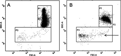
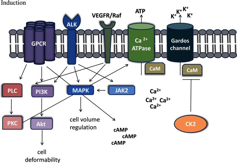
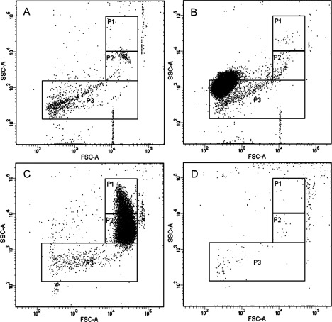
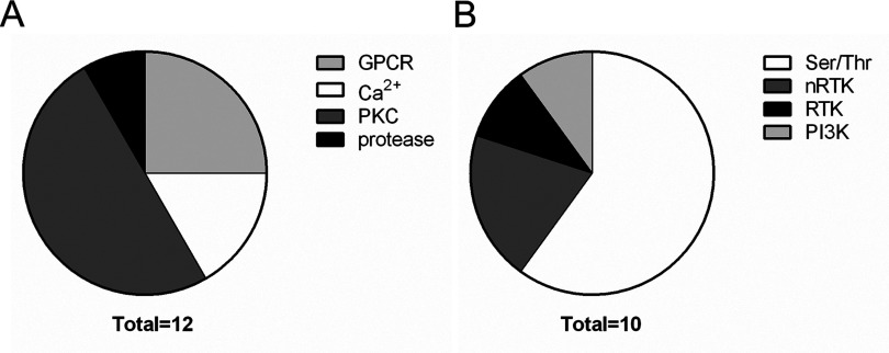
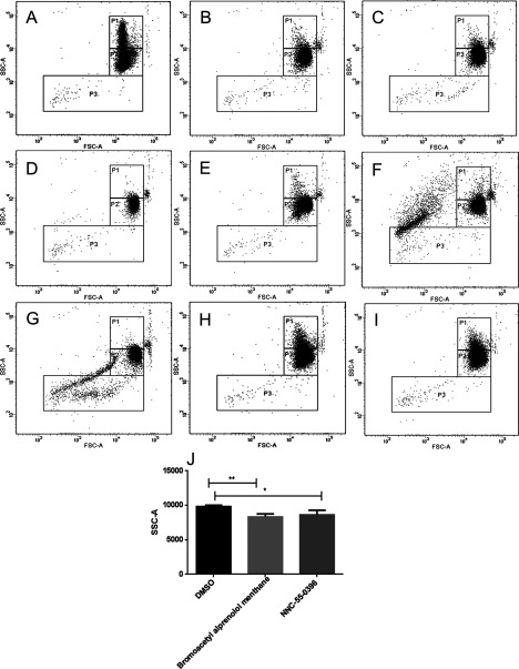
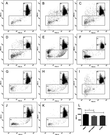
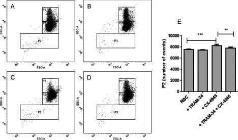
Even though red blood cell (RBC) vesiculation is a well-documented phenomenon, notably in the context of RBC aging and blood transfusion, the exact signalling pathways and kinases involved in this process remain largely unknown. We have established a screening method for RBC vesicle shedding using the Ca(2+) ionophore ionomycin which is a rapid and efficient method to promote vesiculation. In order to identify novel pathways stimulating vesiculation in RBC, we screened two libraries: the Library of Pharmacologically Active Compounds (LOPAC) and the Selleckchem Kinase Inhibitor Library for their effects on RBC from healthy donors. We investigated compounds triggering vesiculation and compounds inhibiting vesiculation induced by ionomycin. We identified 12 LOPAC compounds, nine kinase inhibitors and one kinase activator which induced RBC shrinkage and vesiculation. Thus, we discovered several novel pathways involved in vesiculation including G protein-coupled receptor (GPCR) signalling, the phosphoinositide 3-kinase (PI3K)-Akt (protein kinase B) pathway, the Jak-STAT (Janus kinase-signal transducer and activator of transcription) pathway and the Raf-MEK (mitogen-activated protein kinase kinase)-ERK (extracellular signal-regulated kinase) pathway. Moreover, we demonstrated a link between casein kinase 2 (CK2) and RBC shrinkage via regulation of the Gardos channel activity. In addition, our data showed that inhibition of several kinases with unknown functions in mature RBC, including Alk (anaplastic lymphoma kinase) kinase and vascular endothelial growth factor receptor 2 (VEGFR-2), induced RBC shrinkage and vesiculation.
尽管红细胞(RBC)囊泡化是一个有充分文献记载的现象,特别是在红细胞衰老和输血的背景下,但参与这一过程的确切信号通路和激酶在很大程度上仍不清楚。我们建立了一种使用钙离子载体离子霉素的红细胞囊泡脱落筛选方法,这是一种促进囊泡化的快速有效方法。为了确定刺激红细胞囊泡化的新途径,我们筛选了两个文库:药理活性化合物文库(LOPAC)和Selleckchem激酶抑制剂文库,以研究它们对健康供体红细胞的影响。我们研究了引发囊泡化的化合物以及抑制离子霉素诱导的囊泡化的化合物。我们鉴定出12种LOPAC化合物、9种激酶抑制剂和1种激酶激活剂,它们可诱导红细胞收缩和囊泡化。因此,我们发现了几个参与囊泡化的新途径,包括G蛋白偶联受体(GPCR)信号通路、磷酸肌醇3激酶(PI3K)-Akt(蛋白激酶B)途径、Jak-STAT(Janus激酶-信号转导和转录激活因子)途径以及Raf-MEK(丝裂原活化蛋白激酶激酶)-ERK(细胞外信号调节激酶)途径。此外,我们通过调节加尔多斯通道活性证明了酪蛋白激酶2(CK2)与红细胞收缩之间的联系。此外,我们的数据表明,抑制成熟红细胞中几种功能未知的激酶,包括间变性淋巴瘤激酶(Alk)和血管内皮生长因子受体2(VEGFR-2),可诱导红细胞收缩和囊泡化。