Tilleul Julien, Querques Giuseppe, Capuano Vittorio, Miere Alexandra, Srour Mayer, Souied Eric H
Department of Ophthalmology, Centre Hospitalier Intercommunal de Créteil, University Paris Est Créteil, Créteil, France.
Case Rep Ophthalmol. 2014 Nov 5;5(3):352-6. doi: 10.1159/000369124. eCollection 2014 Sep-Dec.
To report the case of a patient with unilateral idiopathic macular telangiectasia (IMT) associated with type 3 neovascularization.
Observational case report.
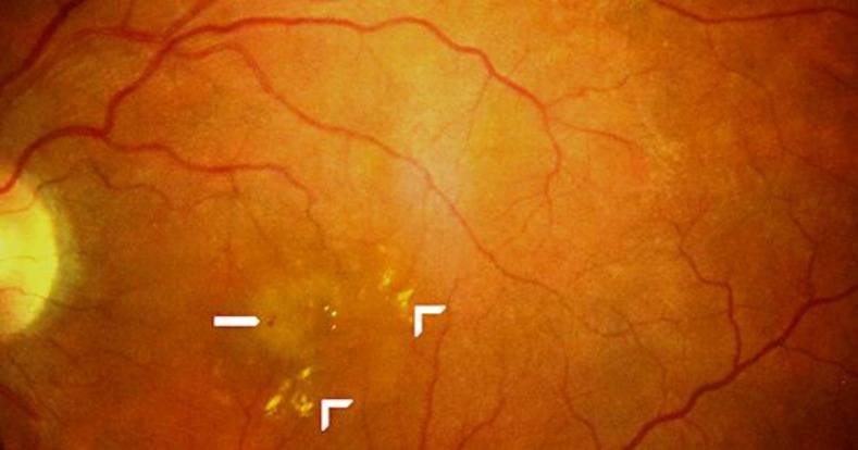
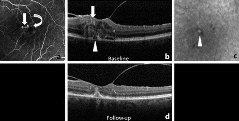
We describe a case of an 85-year-old woman who presented at our department with a gradual vision loss in her left eye (LE). Her best-corrected visual acuity (BCVA) was 20/200 in the LE. Fundus examination showed 2 small hemorrhages located nasally to the LE fovea, as well as lipid exudates. Fluorescein angiography revealed early hyperfluorescence corresponding to the dilated capillaries. Spectral-domain optical coherence tomography (SD-OCT) showed several microaneurysms within the inner retinal layers. Late indocyanine green angiography revealed a focal hyperfluorescence corresponding to a type 3 neovascularization. No signs of IMT or type 3 neovascularization were detected in the right eye. Based on these findings, the patient was diagnosed with type 1 IMT and coincident type 3 neovascularization. The LE was treated with intravitreal anti-vascular endothelial growth factor (anti-VEGF) injections. Twenty-four months later, SD-OCT revealed regression of the exudative signs, and LE BCVA improved to 20/100.
We describe the case of an unusual association between older-onset IMT and type 3 neovascularization, and subsequent regression by anti-VEGF injections. We propose a new IMT subtype called type 1C for this association. Further research must be done in order to establish the pathophysiologic mechanism and likelihood of this association.
报告一例单侧特发性黄斑毛细血管扩张症(IMT)合并3型新生血管形成的病例。
观察性病例报告。
我们描述了一例85岁女性患者,因左眼(LE)视力逐渐下降就诊于我院。其左眼最佳矫正视力(BCVA)为20/200。眼底检查显示左眼黄斑中心凹鼻侧有2处小出血以及脂质渗出。荧光素血管造影显示早期高荧光对应扩张的毛细血管。频域光学相干断层扫描(SD - OCT)显示视网膜内层有多个微动脉瘤。吲哚菁绿血管造影晚期显示一处局灶性高荧光对应3型新生血管形成。右眼未检测到IMT或3型新生血管形成的迹象。基于这些发现,该患者被诊断为1型IMT合并3型新生血管形成。左眼接受了玻璃体内抗血管内皮生长因子(抗VEGF)注射治疗。24个月后,SD - OCT显示渗出性体征消退,左眼BCVA提高到20/100。
我们描述了一例老年发病的IMT与3型新生血管形成之间不寻常的关联,以及随后通过抗VEGF注射实现消退的病例。我们为此关联提出一种新的IMT亚型,称为1C型。必须进行进一步研究以确定这种关联的病理生理机制和可能性。