Gore Jesse, Craven Kelly E, Wilson Julie L, Cote Gregory A, Cheng Monica, Nguyen Hai V, Cramer Harvey M, Sherman Stuart, Korc Murray
Department of Medicine, Indiana University School of Medicine, Indianapolis, IN 46202, USA.
The Melvin and Bren Simon Cancer Center, and The Center for Pancreatic Cancer Research, Indianapolis, IN 46202, USA.
Oncotarget. 2015 Apr 10;6(10):7504-21. doi: 10.18632/oncotarget.3233.
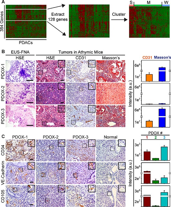
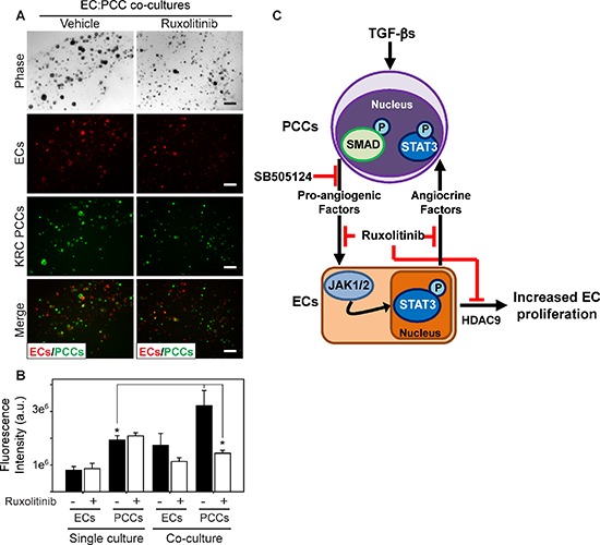
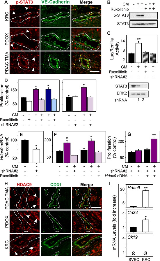
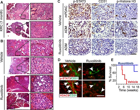
Pancreatic ductal adenocarcinomas (PDACs) overexpress pro-angiogenic factors but are not viewed as vascular. Using data from The Cancer Genome Atlas we demonstrate that a subset of PDACs exhibits a strong pro-angiogenic signature that includes 37 genes, such as HDAC9, that are overexpressed in PDAC arising in KRC mice, which express mutated Kras and lack RB. Moreover, patient-derived orthotopic xenografts can exhibit tumor angiogenesis, whereas conditioned media (CM) from KRC-derived pancreatic cancer cells (PCCs) enhance endothelial cell (EC) growth and migration, and activate canonical TGF-β signaling and STAT3. Inhibition of the type I TGF-β receptor with SB505124 does not alter endothelial activation in vitro, but decreases pro-angiogenic gene expression and suppresses angiogenesis in vivo. Conversely, STAT3 silencing or JAK1-2 inhibition with ruxolitinib blocks CM-enhanced EC proliferation. STAT3 disruption also suppresses endothelial HDAC9 and blocks CM-induced HDAC9 expression, whereas HDAC9 re-expression restores CM-enhanced endothelial proliferation. Moreover, ruxolitinib blocks mitogenic EC/PCC cross-talk, and suppresses endothelial p-STAT3 and HDAC9, and PDAC progression and angiogenesis in vivo, while markedly prolonging survival of KRC mice. Thus, targeting JAK1-2 with ruxolitinib blocks a final pathway that is common to multiple pro-angiogenic factors, suppresses EC-mediated PCC proliferation, and may be useful in PDACs with a strong pro-angiogenic signature.
胰腺导管腺癌(PDAC)过度表达促血管生成因子,但不被视为血管性肿瘤。利用癌症基因组图谱(The Cancer Genome Atlas)的数据,我们证明一部分PDAC表现出强烈的促血管生成特征,该特征包括37个基因,如HDAC9,这些基因在表达突变型Kras且缺乏RB的KRC小鼠所发生的PDAC中过度表达。此外,患者来源的原位异种移植瘤可表现出肿瘤血管生成,而来自KRC来源的胰腺癌细胞(PCC)的条件培养基(CM)可增强内皮细胞(EC)的生长和迁移,并激活经典的TGF-β信号传导和STAT3。用SB505124抑制I型TGF-β受体在体外不会改变内皮细胞的激活,但会降低促血管生成基因的表达并在体内抑制血管生成。相反,用鲁索替尼沉默STAT3或抑制JAK1-2可阻断CM增强的EC增殖。STAT3的破坏还会抑制内皮细胞中的HDAC9并阻断CM诱导的HDAC9表达,而HDAC9的重新表达可恢复CM增强的内皮细胞增殖。此外,鲁索替尼可阻断有丝分裂原性EC/PCC相互作用,并抑制内皮细胞中的p-STAT3和HDAC9,以及体内的PDAC进展和血管生成,同时显著延长KRC小鼠的生存期。因此,用鲁索替尼靶向JAK1-2可阻断多个促血管生成因子共有的最终途径,抑制EC介导的PCC增殖,并且可能对具有强烈促血管生成特征的PDAC有用。