Reed R, Xu C, Liu Y, Gorski J P, Wang Y, Walker M P
Department of Oral and Craniofacial Sciences, School of Dentistry, University of Missouri-Kansas City, MO, United States.
Department of Oral and Craniofacial Sciences, School of Dentistry, University of Missouri-Kansas City, MO, United States; Center of Excellencein Musculoskeletal and Dental Tissues, University of Missouri-Kansas City, MO, United States.
Arch Oral Biol. 2015 May;60(5):690-7. doi: 10.1016/j.archoralbio.2015.02.020. Epub 2015 Feb 27.
To understand radiotherapy-induced dental lesions characterized by enamel loss or delamination near the dentine-enamel junction (DEJ), this study evaluated enamel and dentine nano-mechanical properties and chemical composition before and after simulated oral cancer radiotherapy.
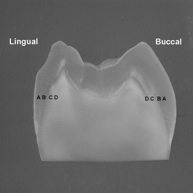
Sections from seven non-carious third molars were exposed to 2 Gy fractions, 5 days/week for 7 weeks for a total of 70 Gy. Nanoindentation was used to evaluate Young's modulus, while Raman microspectroscopy was used to measure protein/mineral ratios, carbonate/phosphate ratios, and phosphate peak width. All measures were completed prior to and following radiation at the same four buccal and lingual sites 500 and 30 μm from the DEJ in enamel and dentine (E-500, E-30, D-30 and D-500).
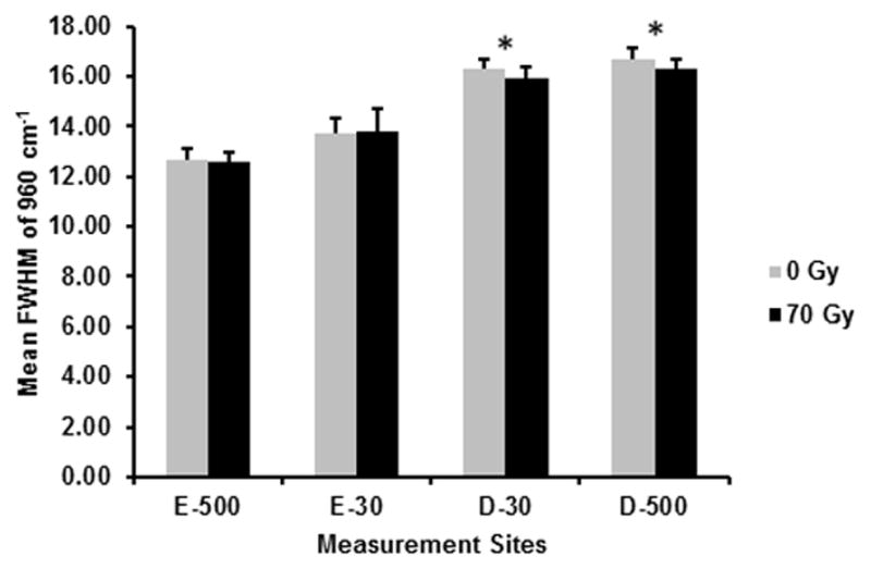
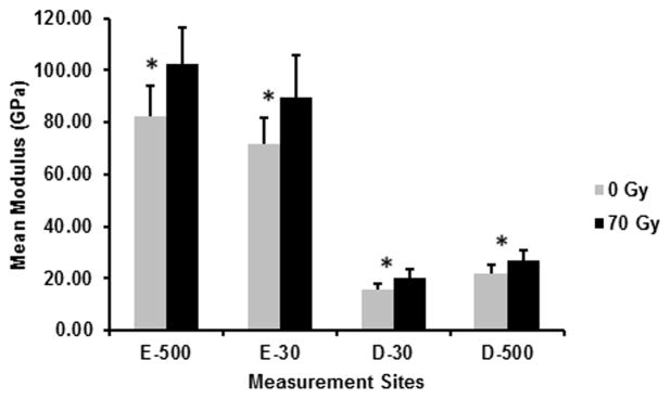
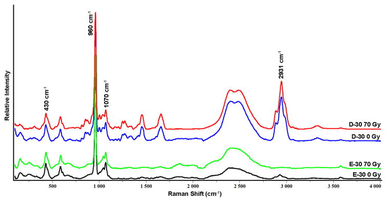
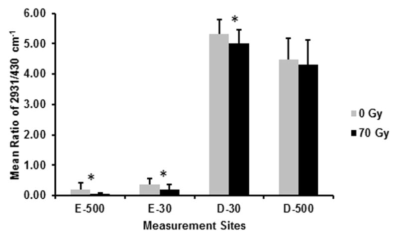
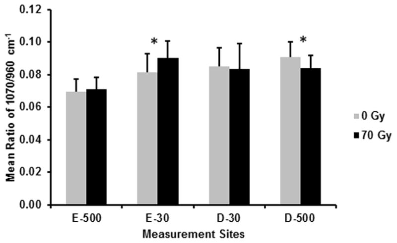
The elastic modulus of enamel and dentine was significantly increased (P ≤ 0.05) following radiation. Based on Raman spectroscopic analysis, there was a significant decrease in the protein to mineral ratio (2931/430 cm(-1)) following radiation at all sites tested except at D-500, while the carbonate to phosphate ratio (1070/960 cm(-1)) increased at E-30 and decreased at D-500. Finally, phosphate peak width as measured by FWHM at 960 cm(-1) significantly decreased at both D-30 and D-500 following radiation.
Simulated radiotherapy produced an increase in the stiffness of enamel and dentine near the DEJ. Increased stiffness is speculated to be the result of the radiation-induced decrease in the protein content, with the percent reduction much greater in the enamel sites. Such changes in mechanical properties and chemical composition could potentially contribute to DEJ biomechanical failure leading to enamel delamination that occurs post-radiotherapy. However, other analyses are required for a better understanding of radiotherapy-induced effects on tooth structure to improve preventive and restorative treatments for oral cancer patients.
为了解以牙釉质在牙本质 - 牙釉质交界(DEJ)附近丧失或分层为特征的放疗诱导性牙齿病变,本研究评估了模拟口腔癌放疗前后牙釉质和牙本质的纳米力学性能及化学成分。
从七颗无龋的第三磨牙获取切片,每周5天,每次接受2 Gy剂量照射,共照射7周,总剂量达70 Gy。采用纳米压痕法评估杨氏模量,同时利用拉曼显微光谱法测量蛋白质/矿物质比率、碳酸盐/磷酸盐比率以及磷酸盐峰宽。所有测量均在距牙釉质和牙本质中DEJ 500和30μm处的相同四个颊侧和舌侧部位在放疗前后完成(E - 500、E - 30、D - 30和D - 500)。
放疗后牙釉质和牙本质的弹性模量显著增加(P≤0.05)。基于拉曼光谱分析,除D - 500外,所有测试部位放疗后蛋白质与矿物质比率(2931/430 cm⁻¹)均显著降低,而碳酸盐与磷酸盐比率(1070/960 cm⁻¹)在E - 30处增加,在D - 500处降低。最后,放疗后D - 30和D - 500处通过半高宽(FWHM)测量的960 cm⁻¹处磷酸盐峰宽均显著降低。
模拟放疗使DEJ附近的牙釉质和牙本质硬度增加。推测硬度增加是放疗诱导蛋白质含量降低的结果,牙釉质部位的降低百分比更大。力学性能和化学成分的此类变化可能会导致放疗后DEJ生物力学失效,进而导致牙釉质分层。然而,需要进行其他分析以更好地理解放疗对牙齿结构的影响,从而改善口腔癌患者的预防和修复治疗。