Thiagarajan Ganesh, Vizcarra Bruno, Bodapudi Venkata, Reed Rachel, Seyedmahmoud Rasoul, Wang Yong, Gorski Jeffrey P, Walker Mary P
a Department of Civil and Mechanical Engineering, School of Computing and Engineering , University of Missouri-Kansas City , Kansas City , MO , USA.
c Center for Excellence in the Study of Dental and Musculoskeletal Tissues , University of Missouri-Kansas City , Kansas City , MO , USA.
Comput Methods Biomech Biomed Engin. 2017 Nov;20(14):1533-1542. doi: 10.1080/10255842.2017.1383401. Epub 2017 Oct 24.
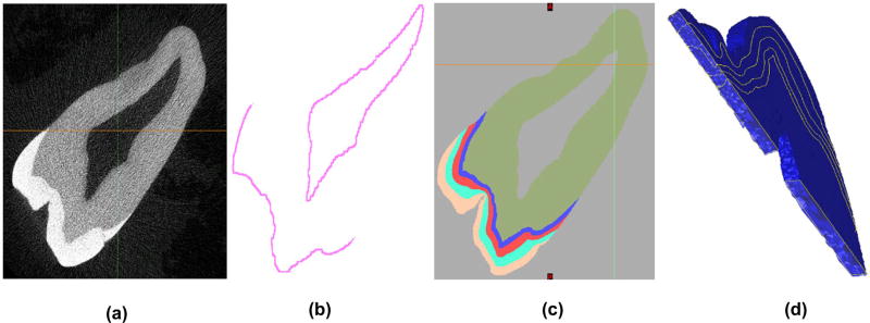
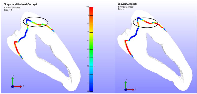
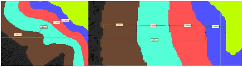
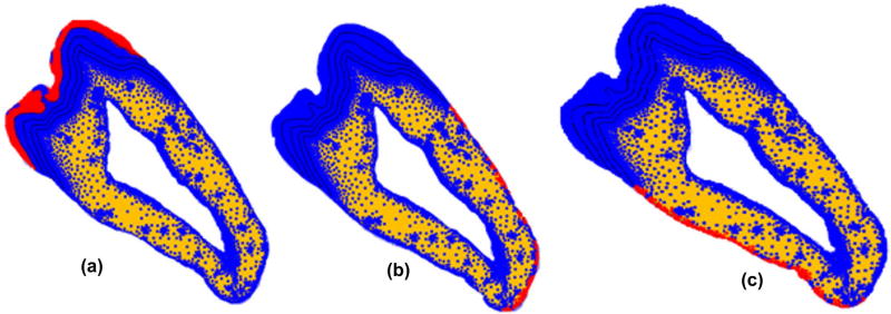
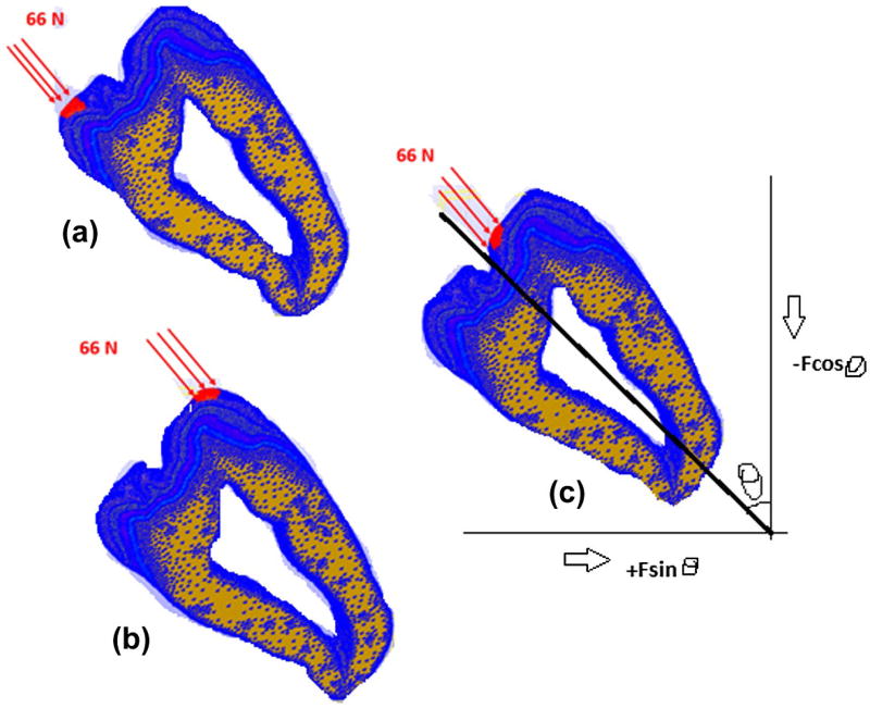
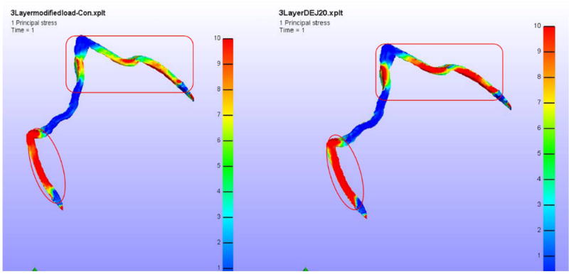
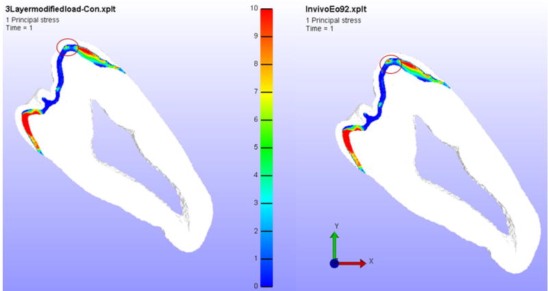
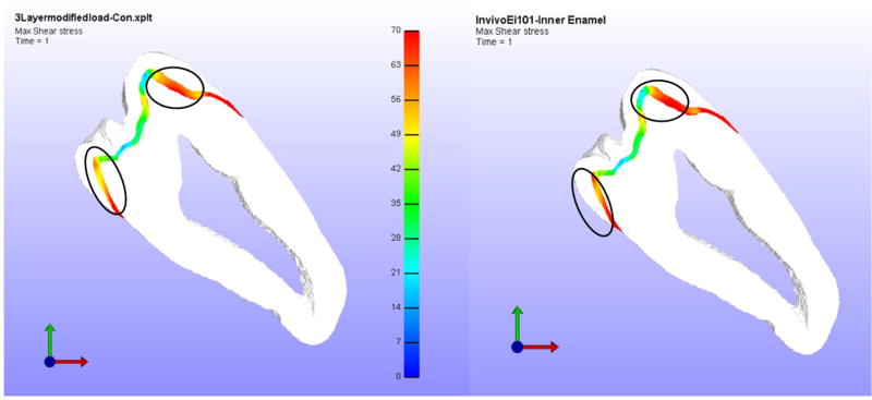
The objectives of this project were to use finite element methods to determine how changes in the elastic modulus due to oral cancer therapeutic radiation alter the distribution of mechanical stresses in teeth and to determine if observed failures in irradiated teeth correlate with changes in mechanical stresses. A thin slice section finite element (FE) model was constructed from micro CT sections of a molar tooth using MIMICS and 3-Matic software. This model divides the tooth into three enamel regions, the dentin-enamel junction (DEJ) and dentin. The enamel elastic modulus was determined in each region using nano indentation for three experimental groups namely - control (non-radiated), in vitro irradiated (simulated radiotherapy following tooth extraction) and in vivo irradiated (extracted subsequent to oral cancer patient radiotherapy) teeth. Physiological loads were applied to the tooth models at the buccal and lingual cusp regions for all three groups (control, in vitro and in vivo). The principal tensile stress and the maximum shear stress were used to compare the results from different groups since it has been observed in previous studies that delamination of enamel from the underlying dentin was one of the major reasons for the failure of teeth following therapeutic radiation. From the FE data, we observed an increase in the principal tensile stress within the inner enamel region of in vivo irradiated teeth (9.97 ± 1.32 MPa) as compared to control/non-irradiated teeth (8.44 ± 1.57 MPa). Our model predicts that failure occurs at the inner enamel/DEJ interface due to extremely high tensile and maximum shear stresses in in vivo irradiated teeth which could be a cause of enamel delamination due to radiotherapy.
本项目的目标是使用有限元方法来确定口腔癌治疗性放疗导致的弹性模量变化如何改变牙齿中的机械应力分布,并确定观察到的受辐照牙齿的损坏是否与机械应力变化相关。使用MIMICS和3-Matic软件,从一颗磨牙的显微CT切片构建了一个薄片截面有限元(FE)模型。该模型将牙齿分为三个牙釉质区域、牙本质-牙釉质交界处(DEJ)和牙本质。使用纳米压痕法测定了三个实验组(即对照组(未辐照)、体外辐照组(拔牙后模拟放疗)和体内辐照组(口腔癌患者放疗后拔牙))牙齿各区域的牙釉质弹性模量。对所有三组(对照组、体外组和体内组)的牙齿模型在颊尖和舌尖区域施加生理载荷。由于在先前的研究中已经观察到牙釉质从其下方牙本质分层是治疗性放疗后牙齿损坏的主要原因之一,因此使用主拉应力和最大剪应力来比较不同组的结果。从有限元数据中,我们观察到与对照组/未辐照牙齿(8.44±1.57MPa)相比,体内辐照牙齿的内牙釉质区域主拉应力增加(9.97±1.32MPa)。我们的模型预测,由于体内辐照牙齿中极高的拉应力和最大剪应力,损坏发生在内牙釉质/DEJ界面,这可能是放疗导致牙釉质分层的一个原因。