Wright Bernice, Parmar Nina, Bozec Laurent, Aguayo Sebastian D, Day Richard M
Applied Biomedical Engineering Group, Division of Medicine, University College London.
Division Biomaterials and Tissue Engineering, UCL Eastman Dental Institute, University College London.
J Biomater Appl. 2015 Aug;30(2):147-59. doi: 10.1177/0885328215577297. Epub 2015 Mar 18.
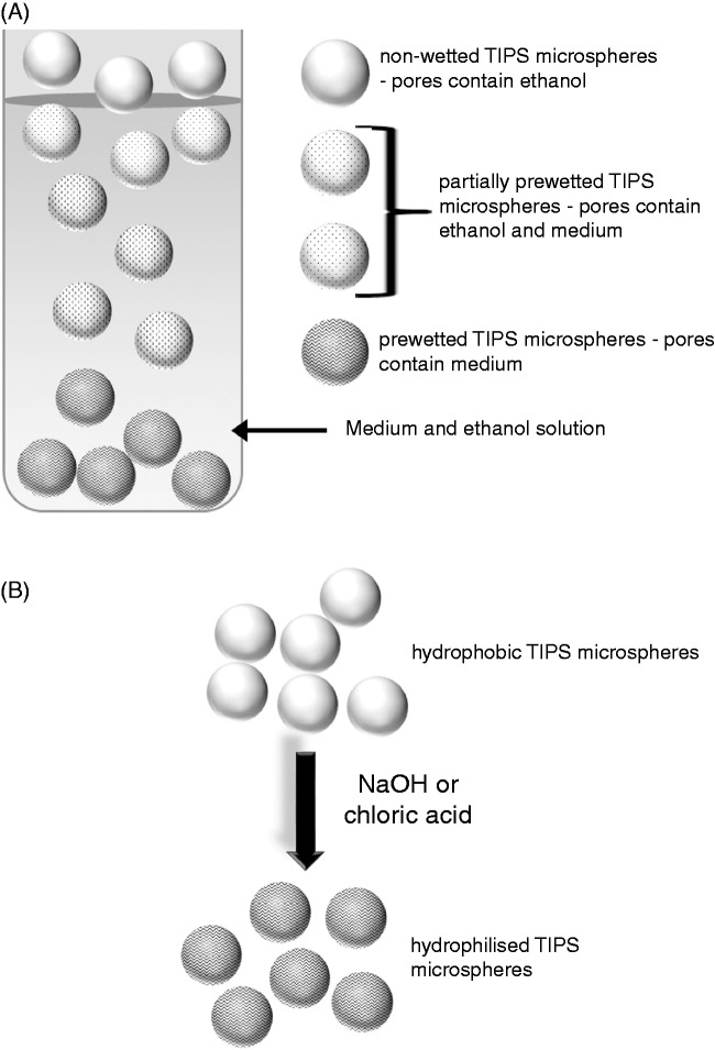
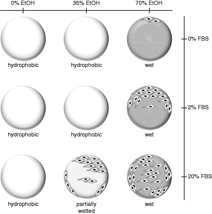
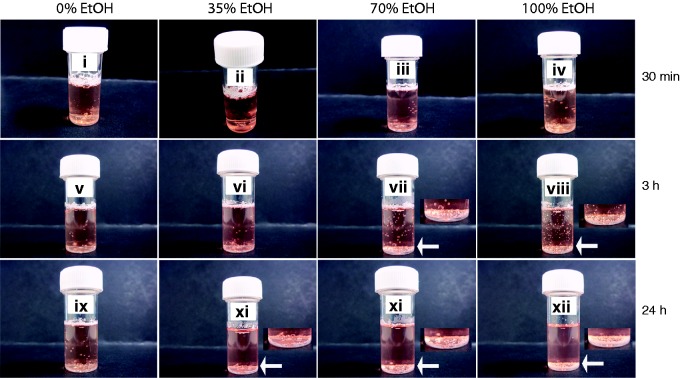
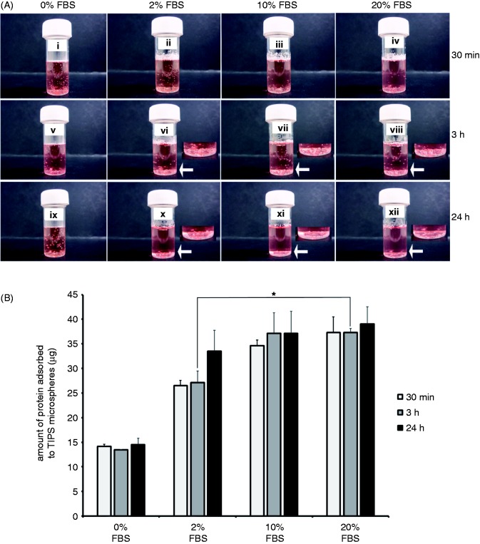
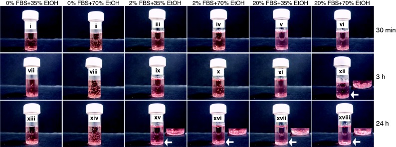
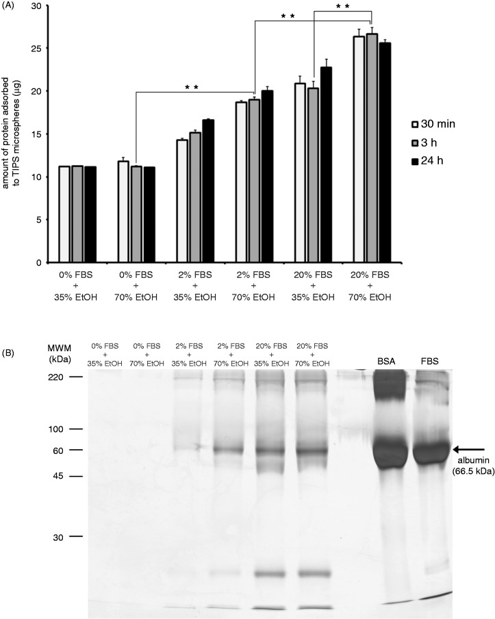
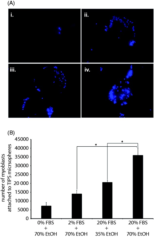
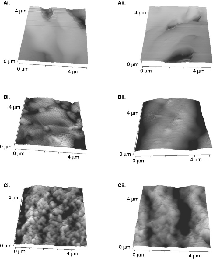
Poly (lactic-co-glycolic) acid microspheres are amenable to a number of biomedical procedures that support delivery of cells, drugs, peptides or genes. Hydrophilisation or wetting of poly (lactic-co-glycolic) acid are an important pre-requisites for attachment of cells and can be achieved via exposure to plasma oxygen or nitrogen, surface hydrolysis with NaOH or chloric acid, immersion in ethanol and water, or prolonged incubation in phosphate buffered saline or cell culture medium. The aim of this study is to develop a simple method for wetting poly (lactic-co-glycolic) acid microspheres for cell delivery applications. A one-step ethanol immersion process that involved addition of serum-supplemented medium and ethanol to PLGA microspheres over 30 min-24 h is described in the present study. This protocol presents a more efficient methodology than conventional two-step wetting procedures. Attachment of human skeletal myoblasts to poly (lactic-co-glycolic) acid microspheres was dependent on extent of wetting, changes in surface topography mediated by ethanol pre-wetting and serum protein adsorption. Ethanol, at 70% (v/v) and 100%, facilitated similar levels of wetting. Wetting with 35% (v/v) ethanol was only achieved after 24 h. Pre-wetting (over 3 h) with 70% (v/v) ethanol allowed significantly greater (p ≤ 0.01) serum protein adsorption to microspheres than wetting with 35% (v/v) ethanol. On serum protein-loaded microspheres, greater numbers of myoblasts attached to constructs wetted with 70% ethanol than those partially wetted with 35% (v/v) ethanol. Microspheres treated with 70% (v/v) ethanol presented a more rugose surface than those treated with 35% (v/v) ethanol, indicating that more efficient myoblast adhesion to the former may be at least partially attributed to differences in surface structure. We conclude that our novel protocol for pre-wetting poly (lactic-co-glycolic) acid microspheres that incorporates biochemical and structural features into this biomaterial can facilitate myoblast delivery for use in clinical settings.
聚乳酸-乙醇酸共聚物微球适用于多种支持细胞、药物、肽或基因递送的生物医学程序。聚乳酸-乙醇酸共聚物的亲水化或湿润是细胞附着的重要先决条件,可通过暴露于等离子体氧气或氮气、用氢氧化钠或氯酸进行表面水解、浸入乙醇和水中,或在磷酸盐缓冲盐水或细胞培养基中长时间孵育来实现。本研究的目的是开发一种用于细胞递送应用的聚乳酸-乙醇酸共聚物微球湿润的简单方法。本研究描述了一种一步乙醇浸入法,即在30分钟至24小时内将补充血清的培养基和乙醇添加到聚乳酸-乙醇酸共聚物微球中。该方案比传统的两步湿润程序提供了一种更有效的方法。人骨骼肌成肌细胞附着于聚乳酸-乙醇酸共聚物微球取决于湿润程度、乙醇预湿润介导的表面形貌变化和血清蛋白吸附。70%(v/v)和100%的乙醇促进了相似程度的湿润。用35%(v/v)乙醇湿润仅在24小时后实现。用70%(v/v)乙醇预湿润(超过3小时)比用35%(v/v)乙醇湿润能使微球上的血清蛋白吸附显著增加(p≤0.01)。在加载血清蛋白的微球上,附着于用70%乙醇湿润的构建体上的成肌细胞数量多于用35%(v/v)乙醇部分湿润的构建体。用70%(v/v)乙醇处理的微球比用35%(v/v)乙醇处理的微球呈现出更粗糙的表面,表明成肌细胞对前者更有效的粘附可能至少部分归因于表面结构的差异。我们得出结论,我们将生化和结构特征纳入这种生物材料的聚乳酸-乙醇酸共聚物微球预湿润新方案可以促进成肌细胞递送,用于临床环境。