• 文献检索
  • 文档翻译
  • 深度研究
  • 学术资讯
  • Suppr Zotero 插件Zotero 插件
  • 邀请有礼
  • 套餐&价格
  • 历史记录
应用&插件
Suppr Zotero 插件Zotero 插件浏览器插件Mac 客户端Windows 客户端微信小程序
定价
高级版会员购买积分包购买API积分包
服务
文献检索文档翻译深度研究API 文档MCP 服务
关于我们
关于 Suppr公司介绍联系我们用户协议隐私条款
关注我们

Suppr 超能文献

核心技术专利:CN118964589B侵权必究
粤ICP备2023148730 号-1Suppr @ 2026

文献检索

告别复杂PubMed语法,用中文像聊天一样搜索,搜遍4000万医学文献。AI智能推荐,让科研检索更轻松。

立即免费搜索

文件翻译

保留排版,准确专业,支持PDF/Word/PPT等文件格式,支持 12+语言互译。

免费翻译文档

深度研究

AI帮你快速写综述,25分钟生成高质量综述,智能提取关键信息,辅助科研写作。

立即免费体验

关于氧化铁纳米颗粒及其对小鼠原代脑细胞不同影响的新发现。

New findings about iron oxide nanoparticles and their different effects on murine primary brain cells.

作者信息

Neubert Jenni, Wagner Susanne, Kiwit Jürgen, Bräuer Anja U, Glumm Jana

机构信息

Institute of Cell Biology and Neurobiology, Center for Anatomy, Charité-Universitaetsmedizin Berlin, Berlin, Germany.

Institute for Radiology, Charité-Universitaetsmedizin Berlin, Berlin, Germany.

出版信息

Int J Nanomedicine. 2015 Mar 13;10:2033-49. doi: 10.2147/IJN.S74404. eCollection 2015.

DOI:10.2147/IJN.S74404
PMID:25792834
原文链接:https://pmc.ncbi.nlm.nih.gov/articles/PMC4364595/
Abstract

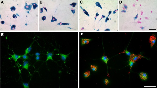
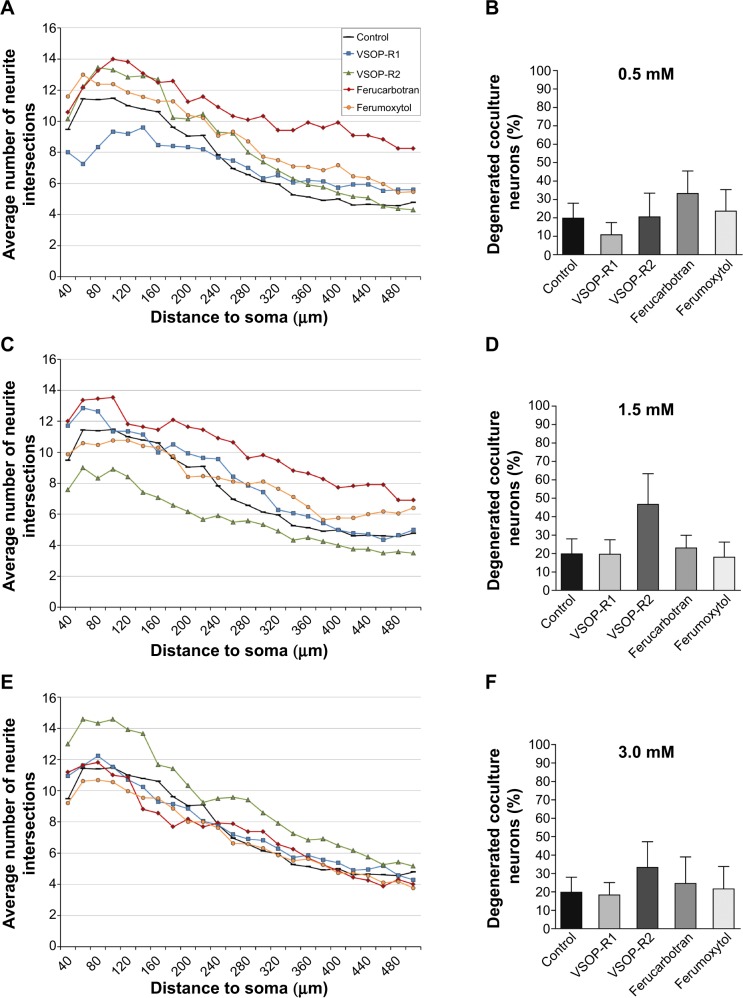
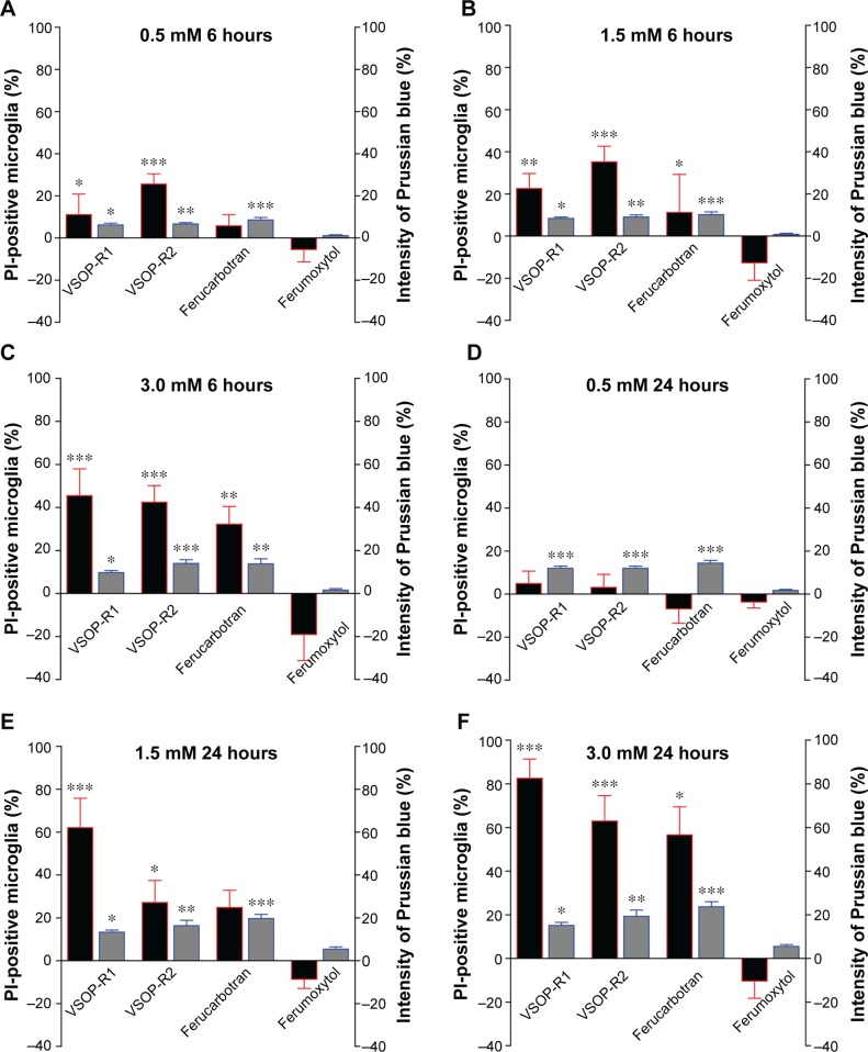
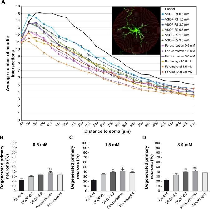
The physicochemical properties of superparamagnetic iron oxide nanoparticles (SPIOs) enable their application in the diagnostics and therapy of central nervous system diseases. However, since crucial information regarding side effects of particle-cell interactions within the central nervous system is still lacking, we investigated the influence of novel very small iron oxide particles or the clinically approved ferucarbotran or ferumoxytol on the vitality and morphology of brain cells. We exposed primary cell cultures of microglia and hippocampal neurons, as well as neuron-glia cocultures to varying concentrations of SPIOs for 6 and/or 24 hours, respectively. Here, we show that SPIO accumulation by microglia and subsequent morphological alterations strongly depend on the respective nanoparticle type. Microglial viability was severely compromised by high SPIO concentrations, except in the case of ferumoxytol. While ferumoxytol did not cause immediate microglial death, it induced severe morphological alterations and increased degeneration of primary neurons. Additionally, primary neurons clearly degenerated after very small iron oxide particle and ferucarbotran exposure. In neuron-glia cocultures, SPIOs rather stimulated the outgrowth of neuronal processes in a concentration- and particle-dependent manner. We conclude that the influence of SPIOs on brain cells not only depends on the particle type but also on the physiological system they are applied to.

摘要

超顺磁性氧化铁纳米颗粒(SPIOs)的物理化学性质使其能够应用于中枢神经系统疾病的诊断和治疗。然而,由于仍缺乏关于中枢神经系统内颗粒与细胞相互作用副作用的关键信息,我们研究了新型超小氧化铁颗粒或临床批准的 ferucarbotran 或 ferumoxytol 对脑细胞活力和形态的影响。我们将小胶质细胞和海马神经元的原代细胞培养物以及神经元 - 胶质细胞共培养物分别暴露于不同浓度的 SPIOs 中 6 小时和/或 24 小时。在此,我们表明小胶质细胞对 SPIO 的摄取以及随后的形态改变强烈依赖于各自的纳米颗粒类型。高浓度的 SPIO 会严重损害小胶质细胞的活力,但 ferumoxytol 除外。虽然 ferumoxytol 不会立即导致小胶质细胞死亡,但它会引起严重的形态改变并增加原代神经元的退化。此外,在暴露于超小氧化铁颗粒和 ferucarbotran 后,原代神经元明显退化。在神经元 - 胶质细胞共培养物中,SPIOs 以浓度和颗粒依赖性方式刺激神经元突起的生长。我们得出结论,SPIOs 对脑细胞的影响不仅取决于颗粒类型,还取决于它们所应用的生理系统。

https://cdn.ncbi.nlm.nih.gov/pmc/blobs/41fc/4364595/07f6e6674c01/ijn-10-2033Fig4.jpg
https://cdn.ncbi.nlm.nih.gov/pmc/blobs/41fc/4364595/b544c5b13a59/ijn-10-2033Fig1.jpg
https://cdn.ncbi.nlm.nih.gov/pmc/blobs/41fc/4364595/dd38f4f31ff3/ijn-10-2033Fig2.jpg
https://cdn.ncbi.nlm.nih.gov/pmc/blobs/41fc/4364595/ebdae0df7929/ijn-10-2033Fig3.jpg
https://cdn.ncbi.nlm.nih.gov/pmc/blobs/41fc/4364595/07f6e6674c01/ijn-10-2033Fig4.jpg

相似文献

1
New findings about iron oxide nanoparticles and their different effects on murine primary brain cells.关于氧化铁纳米颗粒及其对小鼠原代脑细胞不同影响的新发现。
Int J Nanomedicine. 2015 Mar 13;10:2033-49. doi: 10.2147/IJN.S74404. eCollection 2015.
2
Biocompatibility of very small superparamagnetic iron oxide nanoparticles in murine organotypic hippocampal slice cultures and the role of microglia.超小超顺磁性氧化铁纳米颗粒在小鼠海马器官型脑片培养中的生物相容性及小胶质细胞的作用
Int J Nanomedicine. 2017 Feb 27;12:1577-1591. doi: 10.2147/IJN.S127206. eCollection 2017.
3
Lysosomal iron liberation is responsible for the vulnerability of brain microglial cells to iron oxide nanoparticles: comparison with neurons and astrocytes.溶酶体铁释放是脑小胶质细胞对氧化铁纳米颗粒易感性的原因:与神经元和星形胶质细胞的比较。
Nanotoxicology. 2016;10(3):332-42. doi: 10.3109/17435390.2015.1071445. Epub 2015 Aug 18.
4
Cellular magnetic resonance with iron oxide nanoparticles: long-term persistence of SPIO signal in the CNS after transplanted cell death.使用氧化铁纳米颗粒的细胞磁共振成像:移植细胞死亡后,超顺磁性氧化铁(SPIO)信号在中枢神经系统中的长期持续存在。
Nanomedicine (Lond). 2014 Jul;9(10):1457-74. doi: 10.2217/nnm.14.84. Epub 2014 May 13.
5
Cytotoxicity and proliferative capacity impairment induced on human brain cell cultures after short- and long-term exposure to magnetite nanoparticles.短期和长期暴露于磁铁矿纳米颗粒后对人脑细胞培养物诱导产生的细胞毒性和增殖能力损伤。
J Appl Toxicol. 2017 Mar;37(3):361-373. doi: 10.1002/jat.3367. Epub 2016 Aug 2.
6
Endocytotic uptake of iron oxide nanoparticles by cultured brain microglial cells.脑小胶质细胞培养物内吞氧化铁纳米颗粒。
Acta Biomater. 2013 Sep;9(9):8454-65. doi: 10.1016/j.actbio.2013.05.022. Epub 2013 May 31.
7
Comparison of SPIO and USPIO for in vitro labeling of human monocytes: MR detection and cell function.超顺磁性氧化铁(SPIO)和超小超顺磁性氧化铁(USPIO)用于人单核细胞体外标记的比较:磁共振检测与细胞功能
Radiology. 2007 May;243(2):467-74. doi: 10.1148/radiol.2432060120.
8
Cell tagging with clinically approved iron oxides: feasibility and effect of lipofection, particle size, and surface coating on labeling efficiency.使用临床批准的氧化铁进行细胞标记:脂质转染、粒径和表面涂层对标记效率的可行性及影响
Radiology. 2005 Apr;235(1):155-61. doi: 10.1148/radiol.2351040094. Epub 2005 Mar 4.
9
In vitro cytotoxicity of superparamagnetic iron oxide nanoparticles on neuronal and glial cells. Evaluation of nanoparticle interference with viability tests.超顺磁性氧化铁纳米颗粒对神经元和神经胶质细胞的体外细胞毒性。纳米颗粒对活力测试干扰的评估。
J Appl Toxicol. 2016 Mar;36(3):361-72. doi: 10.1002/jat.3213. Epub 2015 Jul 26.
10
Intracellular SPIO labeling of microglia: high field considerations and limitations for MR microscopy.细胞内超顺磁氧化铁标记小胶质细胞:高场考虑因素及磁共振显微镜的局限性。
Contrast Media Mol Imaging. 2012 Mar-Apr;7(2):121-9. doi: 10.1002/cmmi.470.

引用本文的文献

1
Morphology Effect of PEGylated Iron Oxide Nanoparticles on the Enhancement of MRI Contrast Signal in Breast Cancer.聚乙二醇化氧化铁纳米颗粒对乳腺癌磁共振成像对比信号增强的形态学效应
ACS Appl Mater Interfaces. 2025 Aug 6;17(31):44275-44287. doi: 10.1021/acsami.5c12013. Epub 2025 Jul 28.
2
Ferroptosis in radiation-induced brain injury: roles and clinical implications.放射性脑损伤中的铁死亡:作用及临床意义。
Biomed Eng Online. 2024 Sep 11;23(1):93. doi: 10.1186/s12938-024-01288-y.
3
Cellular and Molecular Processes Are Differently Influenced in Primary Neural Cells by Slight Changes in the Physicochemical Properties of Multicore Magnetic Nanoparticles.

本文引用的文献

1
Uptake and metabolism of iron oxide nanoparticles in brain cells.氧化铁纳米颗粒在脑细胞中的摄取与代谢。
Neurochem Res. 2014 Sep;39(9):1648-60. doi: 10.1007/s11064-014-1380-5. Epub 2014 Jul 11.
2
Protein corona composition of superparamagnetic iron oxide nanoparticles with various physico-chemical properties and coatings.具有不同物理化学性质和涂层的超顺磁性氧化铁纳米颗粒的蛋白质冠层组成
Sci Rep. 2014 May 21;4:5020. doi: 10.1038/srep05020.
3
The interplay between iron accumulation, mitochondrial dysfunction, and inflammation during the execution step of neurodegenerative disorders.
多芯磁性纳米粒子理化性质的轻微改变对原代神经细胞的细胞和分子过程有不同的影响。
ACS Appl Mater Interfaces. 2023 Apr 12;15(14):17726-17741. doi: 10.1021/acsami.3c02729. Epub 2023 Mar 28.
4
Magnetic labeling of primary murine monocytes using very small superparamagnetic iron oxide nanoparticles.使用超小超顺磁性氧化铁纳米颗粒对原代小鼠单核细胞进行磁性标记。
Neural Regen Res. 2022 Oct;17(10):2311-2315. doi: 10.4103/1673-5374.336873.
5
Use of Super Paramagnetic Iron Oxide Nanoparticles as Drug Carriers in Brain and Ear: State of the Art and Challenges.超顺磁性氧化铁纳米颗粒在脑和耳中作为药物载体的应用:现状与挑战
Brain Sci. 2021 Mar 11;11(3):358. doi: 10.3390/brainsci11030358.
6
Analysis of the Direct and Indirect Effects of Nanoparticle Exposure on Microglial and Neuronal Cells In Vitro.纳米颗粒暴露对体外小胶质细胞和神经元细胞的直接和间接影响分析。
Int J Mol Sci. 2020 Sep 24;21(19):7030. doi: 10.3390/ijms21197030.
7
Mechanical stimulation of Schwann cells promote peripheral nerve regeneration via extracellular vesicle-mediated transfer of microRNA 23b-3p.施万细胞的机械刺激通过细胞外囊泡介导的 microRNA 23b-3p 转移促进周围神经再生。
Theranostics. 2020 Jul 11;10(20):8974-8995. doi: 10.7150/thno.44912. eCollection 2020.
8
Fluorescence detection of Europium-doped very small superparamagnetic iron oxide nanoparticles in murine hippocampal slice cultures.铕掺杂超小超顺磁性氧化铁纳米颗粒在小鼠海马切片培养物中的荧光检测
Neural Regen Res. 2018 Apr;13(4):637-638. doi: 10.4103/1673-5374.230284.
9
Biocompatibility of very small superparamagnetic iron oxide nanoparticles in murine organotypic hippocampal slice cultures and the role of microglia.超小超顺磁性氧化铁纳米颗粒在小鼠海马器官型脑片培养中的生物相容性及小胶质细胞的作用
Int J Nanomedicine. 2017 Feb 27;12:1577-1591. doi: 10.2147/IJN.S127206. eCollection 2017.
10
The effect of iron nanoparticles on performance of cognitive tasks in rats.铁纳米颗粒对大鼠认知任务表现的影响。
Environ Sci Pollut Res Int. 2017 Mar;24(9):8700-8710. doi: 10.1007/s11356-017-8531-6. Epub 2017 Feb 16.
神经退行性疾病进展阶段铁蓄积、线粒体功能障碍与炎症之间的相互作用。
Front Pharmacol. 2014 Mar 10;5:38. doi: 10.3389/fphar.2014.00038. eCollection 2014.
4
Citrate coated iron oxide nanoparticles with enhanced relaxivity for in vivo magnetic resonance imaging of liver fibrosis.具有增强弛豫率的柠檬酸盐包覆氧化铁纳米颗粒用于肝纤维化的体内磁共振成像。
Colloids Surf B Biointerfaces. 2014 May 1;117:216-24. doi: 10.1016/j.colsurfb.2014.02.034. Epub 2014 Mar 2.
5
Iron deposits in the chronically inflamed central nervous system and contributes to neurodegeneration.慢性炎症的中枢神经系统中的铁沉积,并有助于神经退行性变。
Cell Mol Life Sci. 2014 May;71(9):1607-22. doi: 10.1007/s00018-013-1509-8. Epub 2013 Nov 12.
6
Superparamagnetic iron oxide nanoparticles as MRI contrast agents for non-invasive stem cell labeling and tracking.超顺磁性氧化铁纳米颗粒作为 MRI 对比剂用于非侵入性干细胞标记和示踪。
Theranostics. 2013 Jul 31;3(8):595-615. doi: 10.7150/thno.5366. eCollection 2013.
7
Endocytotic uptake of iron oxide nanoparticles by cultured brain microglial cells.脑小胶质细胞培养物内吞氧化铁纳米颗粒。
Acta Biomater. 2013 Sep;9(9):8454-65. doi: 10.1016/j.actbio.2013.05.022. Epub 2013 May 31.
8
IL-1β and TNF-α induce neurotoxicity through glutamate production: a potential role for neuronal glutaminase.IL-1β 和 TNF-α 通过谷氨酸的产生诱导神经毒性:神经元谷氨酰胺酶的潜在作用。
J Neurochem. 2013 Jun;125(6):897-908. doi: 10.1111/jnc.12263. Epub 2013 May 3.
9
Plasticity-related gene 3 promotes neurite shaft protrusion.可塑性相关基因 3 促进神经突轴突突出。
BMC Neurosci. 2013 Mar 19;14:36. doi: 10.1186/1471-2202-14-36.
10
Characterization of cellular uptake and toxicity of aminosilane-coated iron oxide nanoparticles with different charges in central nervous system-relevant cell culture models.不同表面电荷的氨基硅烷包覆氧化铁纳米颗粒在中枢神经系统相关细胞培养模型中的细胞摄取和毒性特征。
Int J Nanomedicine. 2013;8:961-70. doi: 10.2147/IJN.S39048. Epub 2013 Mar 6.