Libero Lauren E, DeRamus Thomas P, Lahti Adrienne C, Deshpande Gopikrishna, Kana Rajesh K
Department of Psychology, University of Alabama at Birmingham, Birmingham, AL, USA.
Department of Psychiatry and Behavioral Neurobiology, University of Alabama at Birmingham, Birmingham, AL, USA.
Cortex. 2015 May;66:46-59. doi: 10.1016/j.cortex.2015.02.008. Epub 2015 Mar 3.
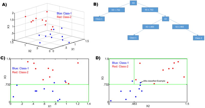
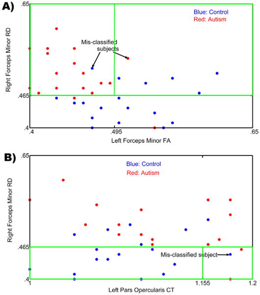
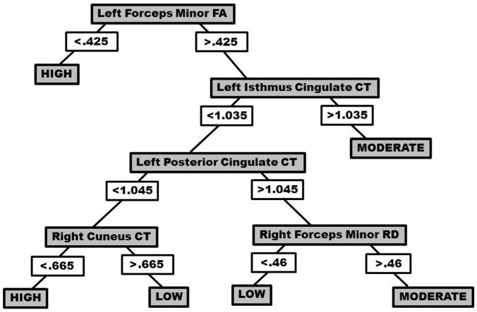
Neuroimaging techniques, such as fMRI, structural MRI, diffusion tensor imaging (DTI), and proton magnetic resonance spectroscopy (1H-MRS) have uncovered evidence for widespread functional and anatomical brain abnormalities in autism spectrum disorder (ASD) suggesting it to be a system-wide neural systems disorder. Nevertheless, most previous studies have focused on examining one index of neuropathology through a single neuroimaging modality, and seldom using multiple modalities to examine the same cohort of individuals. The current study aims to bring together multiple brain imaging modalities (structural MRI, DTI, and 1H-MRS) to investigate the neural architecture in the same set of individuals (19 high-functioning adults with ASD and 18 typically developing (TD) peers). Morphometry analysis revealed increased cortical thickness in ASD participants, relative to typical controls, across the left cingulate, left pars opercularis of the inferior frontal gyrus, left inferior temporal cortex, and right precuneus, and reduced cortical thickness in right cuneus and right precentral gyrus. ASD adults also had reduced fractional anisotropy (FA) and increased radial diffusivity (RD) for two clusters on the forceps minor of the corpus callosum, revealed by DTI analyses. 1H-MRS results showed a reduction in the N-acetylaspartate/Creatine ratio in dorsal anterior cingulate cortex (dACC) in ASD participants. A decision tree classification analysis across the three modalities resulted in classification accuracy of 91.9% with FA, RD, and cortical thickness as key predictors. Examining the same cohort of adults with ASD and their TD peers, this study found alterations in cortical thickness, white matter (WM) connectivity, and neurochemical concentration in ASD. These findings underscore the potential for multimodal imaging to better inform on the neural characteristics most relevant to the disorder.
功能磁共振成像(fMRI)、结构磁共振成像(structural MRI)、扩散张量成像(DTI)和质子磁共振波谱(1H-MRS)等神经成像技术已发现证据,表明自闭症谱系障碍(ASD)存在广泛的功能性和解剖学脑异常,这表明它是一种全系统的神经系统疾病。然而,以前的大多数研究都集中在通过单一神经成像方式检查神经病理学的一个指标,很少使用多种方式检查同一组个体。本研究旨在结合多种脑成像方式(结构MRI、DTI和1H-MRS)来研究同一组个体(19名患有ASD的高功能成年人和18名发育正常(TD)的同龄人)的神经结构。形态学分析显示,与典型对照组相比,ASD参与者的左侧扣带回、左侧额下回岛盖部、左侧颞下皮质和右侧楔前叶的皮质厚度增加,而右侧楔叶和右侧中央前回的皮质厚度减少。DTI分析显示,ASD成年人胼胝体小钳上两个簇的分数各向异性(FA)降低,径向扩散率(RD)增加。1H-MRS结果显示,ASD参与者背侧前扣带回皮质(dACC)的N-乙酰天门冬氨酸/肌酸比值降低。以FA、RD和皮质厚度为关键预测指标的三种成像方式的决策树分类分析得出的分类准确率为91.9%。通过研究同一组患有ASD的成年人及其发育正常的同龄人,本研究发现ASD患者的皮质厚度、白质(WM)连通性和神经化学浓度存在改变。这些发现强调了多模态成像在更好地了解与该疾病最相关的神经特征方面的潜力。