Hyodo Ryota, Komada Tomohiro, Takada Akira, Kawai Hisashi, Ito Shinji, Nishida Yoshihiro, Naganawa Shinji
Department of Radiology, Nagoya University Graduate School of Medicine, Nagoya, Japan.
Department of Radiology, Toyohashi Municipal Hospital, Toyohashi, Japan.
Nagoya J Med Sci. 2015 Feb;77(1-2):167-78.
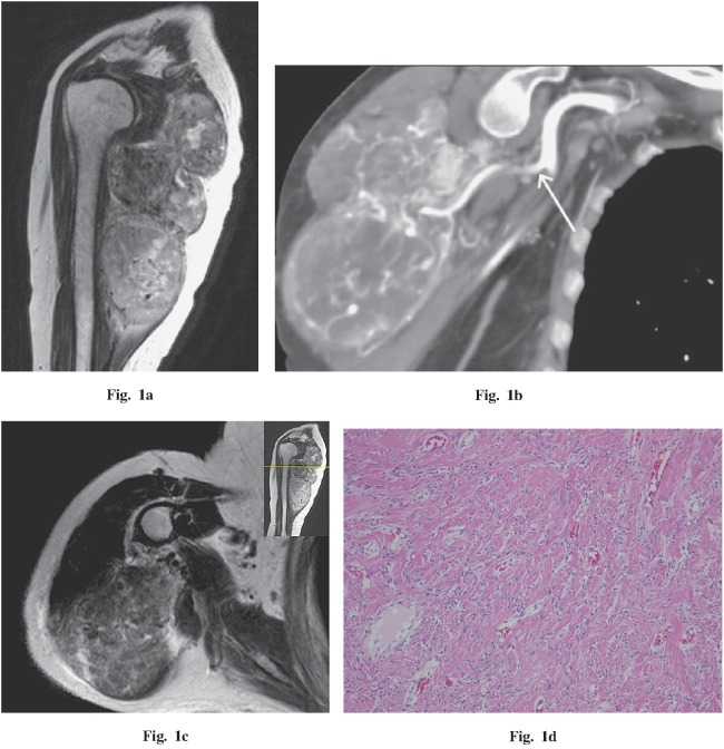
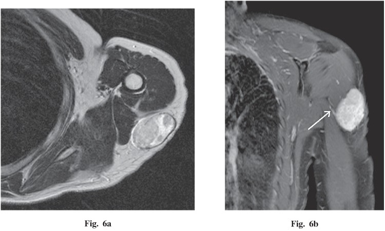
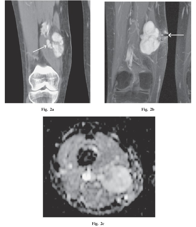
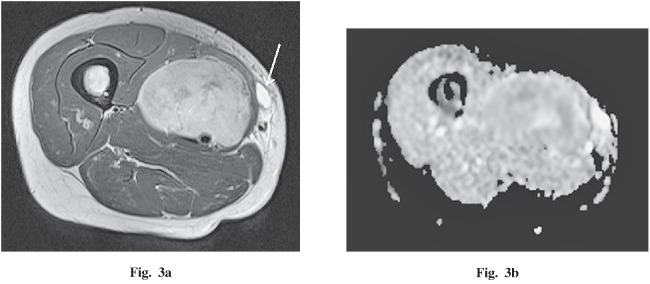
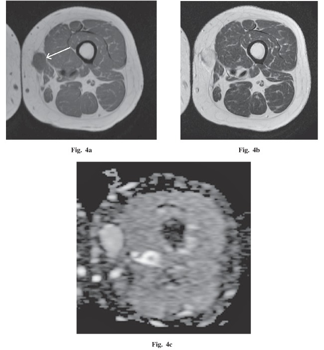
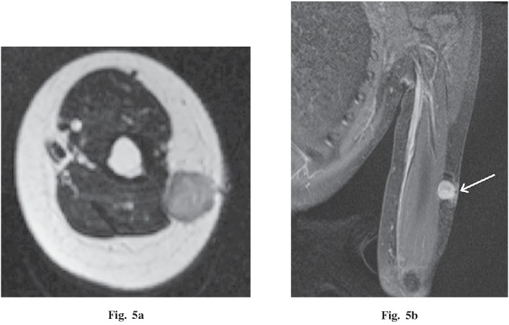
The purpose of this study was to describe the magnetic resonance imaging (MRI) and computed tomography (CT) findings for solitary fibrous tumors (SFTs) in the extremities in correlation with histopathological findings. Between 2006 and 2013, 6 consecutive patients with SFT in an extremity were studied with MRI (6 patients) and CT (4 patients). Diffusion-weighted images were also performed in 3 patients and dynamic contrast-enhanced CT in 2 patients. All 6 tumors were diagnosed after surgical excision, and the pre-surgical imaging findings were correlated with the histopathological findings. As a result, all 6 patients were female, and each had a clearly palpable, well-circumscribed, round or oval mass adjacent to fascia in an extremity, of less than 10 cm maximum diameter in 5 patients. On MRI, the tumors were iso-intense with muscle on T1-weighted image, and appeared heterogeneous and high-intensity on T2-weighted image. After injection of a contrast agent, the tumors demonstrated strong enhancement. A vascular pedicle was detected in 4 patients with tumors having a maximum diameter more than 5 cm. Diffusion-weighted images demonstrated high signal intensities, and apparent diffusion coefficient values were iso to high compared to muscle (from 1.41-2.10×10(-3) mm(2)/s). All the tumors were benign histopathologically and clinically. In 1 patient, the imaging appearance revealed underlying histopathological components, including fibrous-rich, cellular-rich, and myxoid change areas. In conclusion, a SFT in an extremity comprises a well-circumscribed mass adjacent to fascia having a fibrous-dominant area, strong contrast enhancement, and a vascular pedicle.
本研究的目的是描述四肢孤立性纤维性肿瘤(SFT)的磁共振成像(MRI)和计算机断层扫描(CT)表现,并与组织病理学结果相关联。2006年至2013年期间,对6例连续的四肢SFT患者进行了MRI(6例患者)和CT(4例患者)检查。3例患者还进行了扩散加权成像,2例患者进行了动态对比增强CT检查。所有6个肿瘤均在手术切除后确诊,术前影像学表现与组织病理学结果相关。结果显示,所有6例患者均为女性,每例患者在四肢筋膜附近均有一个可清晰触及、边界清楚的圆形或椭圆形肿块,5例患者的肿块最大直径小于10 cm。在MRI上,肿瘤在T1加权像上与肌肉呈等信号,在T2加权像上呈不均匀高信号。注射造影剂后,肿瘤表现为明显强化。4例最大直径超过5 cm的肿瘤患者检测到血管蒂。扩散加权成像显示高信号强度,表观扩散系数值与肌肉相比等至高(1.41 - 2.10×10(-3) mm(2)/s)。所有肿瘤在组织病理学和临床上均为良性。1例患者的影像学表现显示了潜在的组织病理学成分,包括富含纤维、富含细胞和黏液样变区域。总之,四肢SFT表现为筋膜附近边界清楚的肿块,具有纤维为主的区域、明显的对比增强和血管蒂。