• 文献检索
  • 文档翻译
  • 深度研究
  • 学术资讯
  • Suppr Zotero 插件Zotero 插件
  • 邀请有礼
  • 套餐&价格
  • 历史记录
应用&插件
Suppr Zotero 插件Zotero 插件浏览器插件Mac 客户端Windows 客户端微信小程序
定价
高级版会员购买积分包购买API积分包
服务
文献检索文档翻译深度研究API 文档MCP 服务
关于我们
关于 Suppr公司介绍联系我们用户协议隐私条款
关注我们

Suppr 超能文献

核心技术专利:CN118964589B侵权必究
粤ICP备2023148730 号-1Suppr @ 2026

文献检索

告别复杂PubMed语法,用中文像聊天一样搜索,搜遍4000万医学文献。AI智能推荐,让科研检索更轻松。

立即免费搜索

文件翻译

保留排版,准确专业,支持PDF/Word/PPT等文件格式,支持 12+语言互译。

免费翻译文档

深度研究

AI帮你快速写综述,25分钟生成高质量综述,智能提取关键信息,辅助科研写作。

立即免费体验

基于三维锥形束计算机断层扫描的髁突位置和形态按垂直骨骼类型的比较

Three-dimensional cone-beam computed tomography based comparison of condylar position and morphology according to the vertical skeletal pattern.

作者信息

Park In-Young, Kim Ji-Hyun, Park Yang-Ho

机构信息

Department of Orthodontics, Hallym University Sacred Heart Hospital, Anyang, Korea.

Department of Orthodontics, Hallym University Kangdong Sacred Heart Hospital, Seoul, Korea.

出版信息

Korean J Orthod. 2015 Mar;45(2):66-73. doi: 10.4041/kjod.2015.45.2.66. Epub 2015 Mar 19.

DOI:10.4041/kjod.2015.45.2.66
PMID:25798412
原文链接:https://pmc.ncbi.nlm.nih.gov/articles/PMC4367133/
Abstract

OBJECTIVE

To compare condylar position and morphology among different vertical skeletal patterns.

METHODS

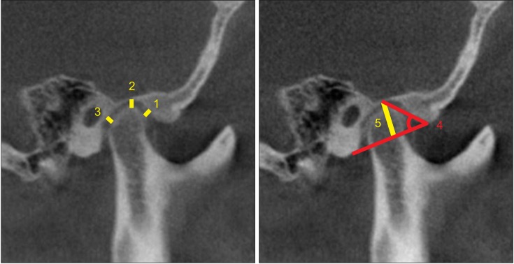
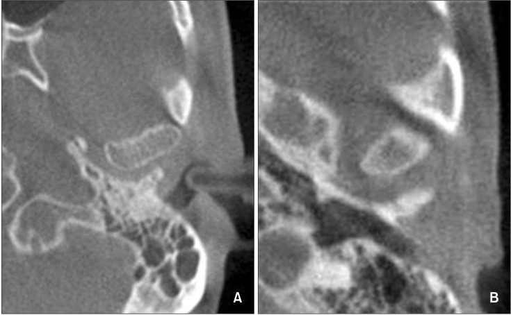
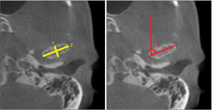
Diagnostic cone-beam computed tomography images of 60 adult patients (120 temporomandibular joints) who visited the orthodontic clinic of Hallym University Sacred Heart Hospital were reviewed. The subjects were divided into three equal groups according to the mandibular plane angle: hypodivergent, normodivergent, and hyperdivergent groups. Morphology of the condyle and mandibular fossa and condylar position were compared among the groups.

RESULTS

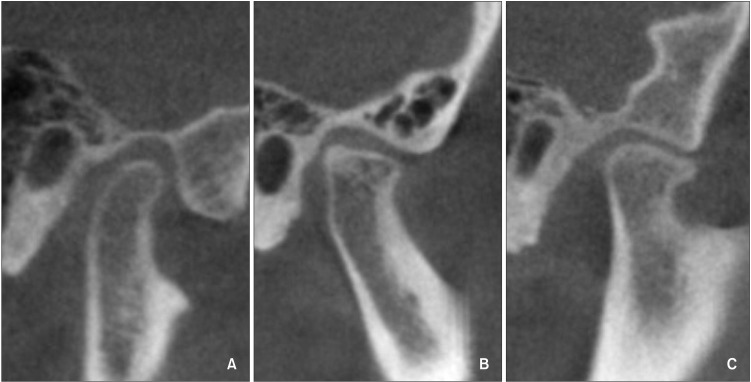
The hypodivergent and hyperdivergent groups showed significant differences in superior joint spaces, antero-posterior condyle width, medio-lateral condyle width, condyle head angle, and condylar shapes.

CONCLUSIONS

Condylar position and morphology vary according to vertical facial morphology. This relationship should be considered for predicting and establishing a proper treatment plan for temporomandibular diseases during orthodontic treatment.

摘要

目的

比较不同垂直骨面型患者髁突的位置及形态。

方法

回顾分析60例成年患者(120个颞下颌关节)的锥形束计算机断层扫描(CBCT)诊断影像,这些患者均就诊于翰林大学圣心医院正畸科。根据下颌平面角将研究对象平均分为三组:低角组、均角组和高角组。比较三组髁突、下颌窝的形态以及髁突位置。

结果

低角组和高角组在上关节间隙、髁突前后径、髁突内外径、髁突头角度及髁突形态方面存在显著差异。

结论

髁突位置及形态随面部垂直形态而异。正畸治疗中预测和制定颞下颌疾病的合理治疗方案时应考虑这种关系。

https://cdn.ncbi.nlm.nih.gov/pmc/blobs/d942/4367133/52916b14a300/kjod-45-66-g004.jpg
https://cdn.ncbi.nlm.nih.gov/pmc/blobs/d942/4367133/2364d6211b5c/kjod-45-66-g001.jpg
https://cdn.ncbi.nlm.nih.gov/pmc/blobs/d942/4367133/163311aed9c0/kjod-45-66-g002.jpg
https://cdn.ncbi.nlm.nih.gov/pmc/blobs/d942/4367133/b85d9e6dc8bc/kjod-45-66-g003.jpg
https://cdn.ncbi.nlm.nih.gov/pmc/blobs/d942/4367133/52916b14a300/kjod-45-66-g004.jpg

相似文献

1
Three-dimensional cone-beam computed tomography based comparison of condylar position and morphology according to the vertical skeletal pattern.基于三维锥形束计算机断层扫描的髁突位置和形态按垂直骨骼类型的比较
Korean J Orthod. 2015 Mar;45(2):66-73. doi: 10.4041/kjod.2015.45.2.66. Epub 2015 Mar 19.
2
[Cone-beam CT evaluation of temporomandibular joint in skeletal class Ⅱ female adolescents with different vertical patterns].[锥形束CT对不同垂直骨面型Ⅱ类女性青少年颞下颌关节的评估]
Beijing Da Xue Xue Bao Yi Xue Ban. 2020 Dec 29;53(1):109-119. doi: 10.19723/j.issn.1671-167X.2021.01.017.
3
Comparison of temporomandibular joints in relation to ages and vertical facial types in skeletal class II female patients: a multiple-cross-sectional study.比较不同年龄和垂直面型的骨性 II 类女性患者颞下颌关节:一项多横断面研究。
BMC Oral Health. 2024 Apr 17;24(1):467. doi: 10.1186/s12903-024-04219-4.
4
Comprehensive three-dimensional positional and morphological assessment of the temporomandibular joint in skeletal Class II patients with mandibular retrognathism in different vertical skeletal patterns.不同垂直骨面型下颌后缩的骨性 II 类错(牙合)患者颞下颌关节的全面三维位置和形态评估。
BMC Oral Health. 2022 Apr 28;22(1):149. doi: 10.1186/s12903-022-02174-6.
5
Three-Dimensional Assessment of Temporomandibular Joint Morphology and Facial Asymmetry in Individuals with Different Vertical Skeletal Growth Patterns.不同垂直骨生长型个体的颞下颌关节形态和面部不对称的三维评估。
Int J Environ Res Public Health. 2023 Jan 12;20(2):1437. doi: 10.3390/ijerph20021437.
6
Differences in mandibular condyle and glenoid fossa morphology in relation to vertical and sagittal skeletal patterns: A cone-beam computed tomography study.下颌髁突与关节窝形态在垂直和矢状向骨骼类型方面的差异:一项锥形束计算机断层扫描研究。
Korean J Orthod. 2021 Mar 25;51(2):126-134. doi: 10.4041/kjod.2021.51.2.126.
7
[Cone-beam CT analysis of vertical control of mandible and changes of temporomandibular joint in adult patients with skeletal class Ⅱ malocclusion with high angle].[成人骨性Ⅱ类高角错牙合患者下颌垂直向控制及颞下颌关节变化的锥形束CT分析]
Zhonghua Kou Qiang Yi Xue Za Zhi. 2022 Nov 9;57(11):1147-1155. doi: 10.3760/cma.j.cn112144-20220301-00086.
8
Comprehensive 3D analysis of condylar morphology in adults with different skeletal patterns - a cross-sectional study.不同骨骼类型成年人髁突形态的综合 3D 分析——一项横断面研究。
Head Face Med. 2020 Nov 30;16(1):33. doi: 10.1186/s13005-020-00245-z.
9
Temporomandibular condylar morphology in diverse maxillary-mandibular skeletal patterns: A 3-dimensional cone-beam computed tomography study.不同上颌-下颌骨骼形态中的颞下颌髁形态:一项三维锥形束 CT 研究。
J Am Dent Assoc. 2018 Jul;149(7):589-598. doi: 10.1016/j.adaj.2018.02.016. Epub 2018 Apr 11.
10
Association between condylar position and vertical skeletal craniofacial morphology: A cone beam computed tomography study.髁突位置与垂直向骨骼颅面形态的相关性:一项锥形束计算机断层扫描研究。
Int Orthod. 2017 Dec;15(4):740-751. doi: 10.1016/j.ortho.2017.09.007. Epub 2017 Oct 28.

引用本文的文献

1
Positional and dimensional osseous characteristics of the temporomandibular joint in female patients with skeletal class III malocclusion and disc displacement, with and without reduction.伴有或不伴有可复性的骨骼Ⅲ类错牙合畸形及盘移位女性患者颞下颌关节的位置和尺寸骨性特征
Front Oral Health. 2025 May 15;6:1572305. doi: 10.3389/froh.2025.1572305. eCollection 2025.
2
Position and Dimensions of the Mandibular Condyle in Various Anterior-Posterior Skeletal Patterns: A CBCT Imaging Study in a Sample of Iranian People.不同前后骨骼模式下下颌髁突的位置与尺寸:一项针对伊朗人群样本的CBCT影像学研究
Int J Dent. 2024 Dec 27;2024:5895594. doi: 10.1155/ijod/5895594. eCollection 2024.
3

本文引用的文献

1
Comparison of condylar displacement between three biotypological facial groups by using mounted models and a mandibular position indicator.使用咬合模型和下颌位置指示器比较三种生物类型面部组之间的髁突移位情况。
Korean J Orthod. 2014 Nov;44(6):312-9. doi: 10.4041/kjod.2014.44.6.312. Epub 2014 Nov 24.
2
Correlation between articular eminence steepness measured with cone-beam computed tomography and clinical dysfunction index in patients with temporomandibular joint dysfunction.关节结节陡峭度与颞下颌关节功能紊乱患者临床功能障碍指数的相关性:锥形束 CT 测量。
Oral Surg Oral Med Oral Pathol Oral Radiol. 2013 Jul;116(1):91-7. doi: 10.1016/j.oooo.2013.04.001.
3
Effect of maxillary molars distalization using clear aligners and fixed orthodontic appliances on the positional and dimensional temporomandibular joint parameters: a three-dimensional comparative study.
使用透明牙套和固定正畸器具远移上颌磨牙对颞下颌关节位置和维度参数的影响:一项三维对比研究。
BMC Oral Health. 2024 Nov 13;24(1):1372. doi: 10.1186/s12903-024-05148-y.
4
Prevalence of condylar morphological changes in individuals with class II malocclusion.安氏II类错牙合个体髁突形态变化的患病率。
Braz Oral Res. 2024 Jul 12;38:e060. doi: 10.1590/1807-3107bor-2024.vol38.0060. eCollection 2024.
5
Comparison of temporomandibular joints in relation to ages and vertical facial types in skeletal class II female patients: a multiple-cross-sectional study.比较不同年龄和垂直面型的骨性 II 类女性患者颞下颌关节:一项多横断面研究。
BMC Oral Health. 2024 Apr 17;24(1):467. doi: 10.1186/s12903-024-04219-4.
6
Volumetric comparison of mandibular condyles and mandibles in the different skeletal classes in the Saudi population.沙特人群不同骨骼类型下颌髁突和下颌骨的体积比较。
PeerJ. 2024 Jan 4;12:e16750. doi: 10.7717/peerj.16750. eCollection 2024.
7
Correlation between facial asymmetry of skeletal class III jaw relationship and morphology of the temporomandibular joint: A cone beam computed tomography study.骨性III类错颌关系面部不对称与颞下颌关节形态的相关性:一项锥形束计算机断层扫描研究。
J Dent Sci. 2023 Jul;18(3):1031-1041. doi: 10.1016/j.jds.2022.11.031. Epub 2022 Dec 12.
8
Dimensional and positional temporomandibular joint osseous characteristics in normodivergent facial patterns with and without temporomandibular disorders.正常颅面型伴或不伴颞下颌关节紊乱患者的颞下颌关节骨性特征的三维位置分析。
Clin Oral Investig. 2023 Sep;27(9):5011-5020. doi: 10.1007/s00784-023-05120-0. Epub 2023 Jun 26.
9
Quantitative and qualitative condylar changes following stabilization splint therapy in patients with temporomandibular joint disorders.髁突定量和定性变化与颞下颌关节紊乱患者稳定夹板治疗的相关性。
Clin Oral Investig. 2023 May;27(5):2299-2310. doi: 10.1007/s00784-023-04963-x. Epub 2023 Apr 11.
10
Comprehensive positional and morphological assessments of the temporomandibular joint in adolescents with skeletal Class III malocclusion: a retrospective CBCT study.青少年骨性 III 类错颌患者颞下颌关节的全面位置和形态评估:一项回顾性 CBCT 研究。
BMC Oral Health. 2023 Feb 7;23(1):78. doi: 10.1186/s12903-023-02788-4.
Assessing joint space and condylar position in the people with normal function of temporomandibular joint with cone-beam computed tomography.
使用锥形束计算机断层扫描评估颞下颌关节功能正常人群的关节间隙和髁突位置。
Dent Res J (Isfahan). 2012 Sep;9(5):607-12. doi: 10.4103/1735-3327.104881.
4
Condylar bony changes in patients with temporomandibular disorders: a CBCT study.颞下颌关节紊乱病患者髁突骨质改变:一项锥形束计算机断层扫描研究
Imaging Sci Dent. 2012 Dec;42(4):249-53. doi: 10.5624/isd.2012.42.4.249. Epub 2012 Dec 23.
5
Disc displacement within the human temporomandibular joint: a systematic review of a 'noisy annoyance'.人类颞下颌关节内的盘移位:对“噪音困扰”的系统综述
J Oral Rehabil. 2013 Feb;40(2):139-58. doi: 10.1111/joor.12016. Epub 2012 Nov 30.
6
Measurement accuracy of temporomandibular joint space in Promax 3-dimensional cone-beam computerized tomography images.髁突关节间隙在 Promax 三维锥形束 CT 图像中的测量准确性。
Oral Surg Oral Med Oral Pathol Oral Radiol. 2012 Jul;114(1):112-7. doi: 10.1016/j.oooo.2011.11.020. Epub 2012 Apr 6.
7
Do skeletal cephalometric characteristics correlate with condylar volume, surface and shape? A 3D analysis.颅面骨骼的形态特征是否与髁突体积、表面和形态相关?一项三维分析。
Head Face Med. 2012 May 15;8:15. doi: 10.1186/1746-160X-8-15.
8
Skeletal differences in patients with temporomandibular joint disc displacement according to sagittal jaw relationship.根据矢状颌关系,颞下颌关节盘移位患者的骨骼差异。
J Oral Maxillofac Surg. 2012 May;70(5):e349-60. doi: 10.1016/j.joms.2011.08.027. Epub 2011 Dec 9.
9
Occlusal force, electromyographic activity of masticatory muscles and mandibular flexure of subjects with different facial types.不同面型受试者的咬合力、咀嚼肌肌电图活动和下颌弯曲。
J Appl Oral Sci. 2011 Aug;19(4):343-9. doi: 10.1590/s1678-77572011005000008. Epub 2011 Jun 3.
10
Computed tomography evaluation of the temporomandibular joint in Class II Division 1 and Class III malocclusion patients: condylar symmetry and condyle-fossa relationship.安氏II类1分类和安氏III类错牙合患者颞下颌关节的计算机断层扫描评估:髁突对称性及髁突-关节窝关系
Am J Orthod Dentofacial Orthop. 2009 Aug;136(2):199-206. doi: 10.1016/j.ajodo.2007.07.033.