Spitale Robert C, Flynn Ryan A, Zhang Qiangfeng Cliff, Crisalli Pete, Lee Byron, Jung Jong-Wha, Kuchelmeister Hannes Y, Batista Pedro J, Torre Eduardo A, Kool Eric T, Chang Howard Y
Howard Hughes Medical Institute and Program in Epithelial Biology, Stanford University School of Medicine, Stanford, California 94305, USA.
Department of Chemistry, Stanford University, Stanford, California 94305, USA.
Nature. 2015 Mar 26;519(7544):486-90. doi: 10.1038/nature14263. Epub 2015 Mar 18.
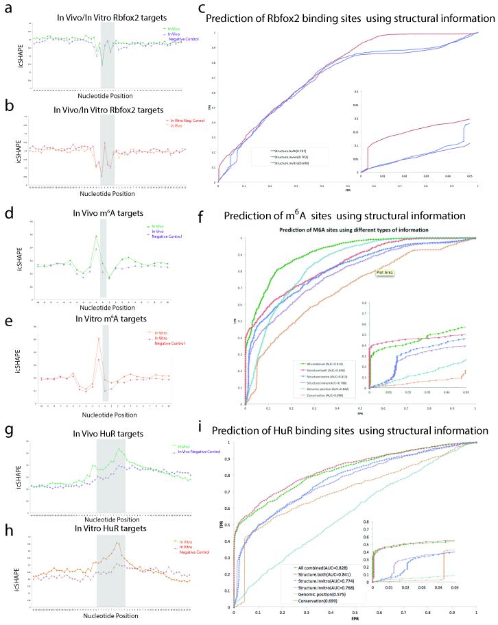
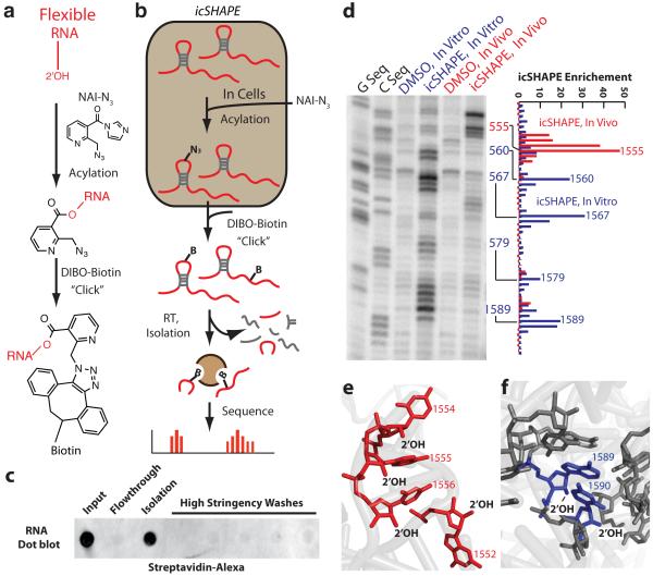
Visualizing the physical basis for molecular behaviour inside living cells is a great challenge for biology. RNAs are central to biological regulation, and the ability of RNA to adopt specific structures intimately controls every step of the gene expression program. However, our understanding of physiological RNA structures is limited; current in vivo RNA structure profiles include only two of the four nucleotides that make up RNA. Here we present a novel biochemical approach, in vivo click selective 2'-hydroxyl acylation and profiling experiment (icSHAPE), which enables the first global view, to our knowledge, of RNA secondary structures in living cells for all four bases. icSHAPE of the mouse embryonic stem cell transcriptome versus purified RNA folded in vitro shows that the structural dynamics of RNA in the cellular environment distinguish different classes of RNAs and regulatory elements. Structural signatures at translational start sites and ribosome pause sites are conserved from in vitro conditions, suggesting that these RNA elements are programmed by sequence. In contrast, focal structural rearrangements in vivo reveal precise interfaces of RNA with RNA-binding proteins or RNA-modification sites that are consistent with atomic-resolution structural data. Such dynamic structural footprints enable accurate prediction of RNA-protein interactions and N(6)-methyladenosine (m(6)A) modification genome wide. These results open the door for structural genomics of RNA in living cells and reveal key physiological structures controlling gene expression.
可视化活细胞内分子行为的物理基础对生物学而言是一项巨大挑战。RNA是生物调控的核心,RNA形成特定结构的能力密切控制着基因表达程序的每一步。然而,我们对生理RNA结构的了解有限;目前的体内RNA结构图谱仅涵盖构成RNA的四种核苷酸中的两种。在此,我们提出一种新的生化方法——体内点击选择性2'-羟基酰化和分析实验(icSHAPE),据我们所知,该方法能够首次全面观察活细胞中所有四种碱基的RNA二级结构。对小鼠胚胎干细胞转录组进行icSHAPE分析,并与体外折叠的纯化RNA进行对比,结果表明细胞环境中RNA的结构动态能够区分不同类别的RNA和调控元件。翻译起始位点和核糖体暂停位点的结构特征在体外条件下是保守的,这表明这些RNA元件由序列编程。相比之下,体内的局部结构重排揭示了RNA与RNA结合蛋白或RNA修饰位点的精确界面,这与原子分辨率的结构数据一致。这种动态结构印记能够在全基因组范围内准确预测RNA-蛋白质相互作用和N(6)-甲基腺苷(m(6)A)修饰。这些结果为活细胞中RNA的结构基因组学打开了大门,并揭示了控制基因表达的关键生理结构。