Mengistu Meron, Ray Krishanu, Lewis George K, DeVico Anthony L
The Institute of Human Virology of the University of Maryland School of Medicine, Baltimore, Maryland, United States of America.
Center for Fluorescence Spectroscopy of the University of Maryland School of Medicine, Baltimore, Maryland, United States of America.
PLoS Pathog. 2015 Mar 25;11(3):e1004772. doi: 10.1371/journal.ppat.1004772. eCollection 2015 Mar.
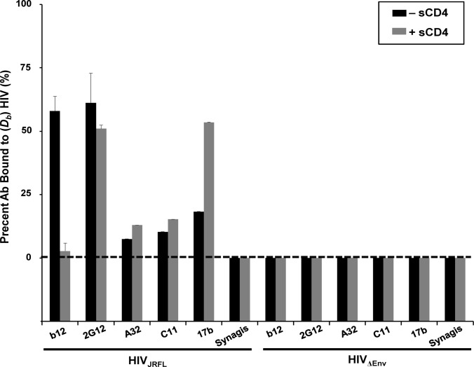
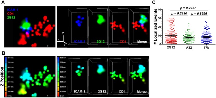
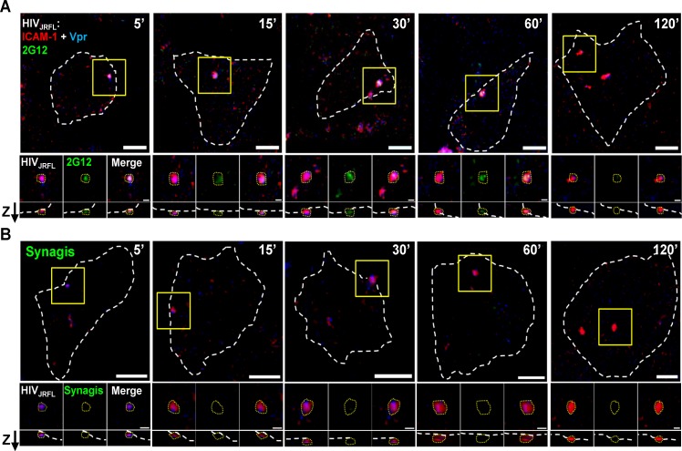
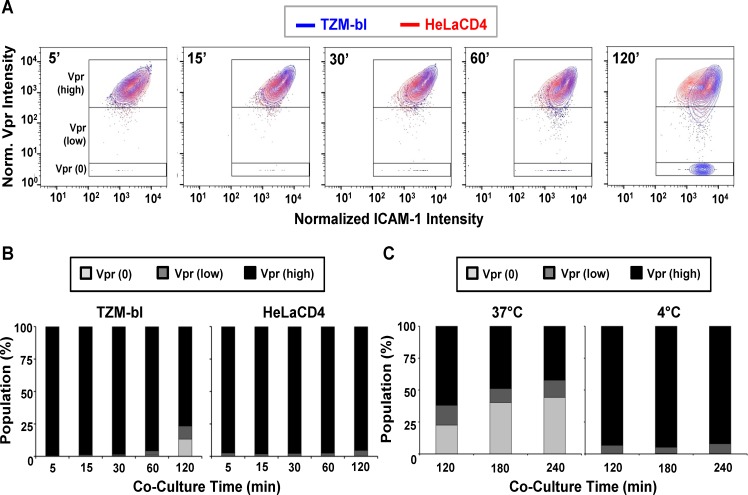
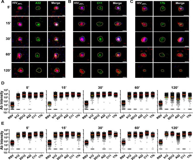
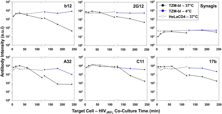
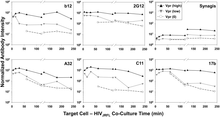
The HIV-1 envelope glycoprotein, gp120, undergoes multiple molecular interactions and structural rearrangements during the course of host cell attachment and viral entry, which are being increasingly defined at the atomic level using isolated proteins. In comparison, antigenic markers of these dynamic changes are essentially unknown for single HIV-1 particles bound to target cells. Such markers should indicate how neutralizing and/or non-neutralizing antibodies might interdict infection by either blocking infection or sensitizing host cells for elimination by Fc-mediated effector function. Here we address this deficit by imaging fluorescently labeled CCR5-tropic HIV-1 pseudoviruses using confocal and superresolution microscopy to track the exposure of neutralizing and non-neutralizing epitopes as they appear on single HIV-1 particles bound to target cells. Epitope exposure was followed under conditions permissive or non-permissive for viral entry to delimit changes associated with virion binding from those associated with post-attachment events. We find that a previously unexpected array of gp120 epitopes is exposed rapidly upon target cell binding. This array comprises both neutralizing and non-neutralizing epitopes, the latter being hidden on free virions yet capable of serving as potent targets for Fc-mediated effector function. Under non-permissive conditions for viral entry, both neutralizing and non-neutralizing epitope exposures were relatively static over time for the majority of bound virions. Under entry-permissive conditions, epitope exposure patterns changed over time on subsets of virions that exhibited concurrent variations in virion contents. These studies reveal that bound virions are distinguished by a broad array of both neutralizing and non-neutralizing gp120 epitopes that potentially sensitize a freshly engaged target cell for destruction by Fc-mediated effector function and/or for direct neutralization at a post-binding step. The elucidation of these epitope exposure patterns during viral entry will help clarify antibody-mediated inhibition of HIV-1 as it is measured in vitro and in vivo.
HIV-1包膜糖蛋白gp120在宿主细胞附着和病毒进入过程中会经历多种分子相互作用和结构重排,利用分离出的蛋白质在原子水平上对这些过程的定义越来越清晰。相比之下,对于与靶细胞结合的单个HIV-1颗粒,这些动态变化的抗原标志物基本上是未知的。此类标志物应能表明中和抗体和/或非中和抗体如何通过阻断感染或使宿主细胞对Fc介导的效应功能敏感以被清除来阻断感染。在这里,我们通过共聚焦显微镜和超分辨率显微镜对荧光标记的CCR5嗜性HIV-1假病毒进行成像,以追踪中和及非中和表位在与靶细胞结合的单个HIV-1颗粒上出现时的暴露情况,从而解决这一不足。在允许或不允许病毒进入的条件下追踪表位暴露情况,以区分与病毒体结合相关的变化和与附着后事件相关的变化。我们发现,与靶细胞结合后,一系列先前未预料到的gp120表位会迅速暴露。该系列包括中和表位和非中和表位,后者在游离病毒体上是隐藏的,但能够作为Fc介导的效应功能的有效靶点。在不允许病毒进入的条件下,对于大多数结合的病毒体,中和及非中和表位暴露随时间相对稳定。在允许进入的条件下,对于病毒体内容物同时发生变化的病毒体亚群,表位暴露模式随时间发生变化。这些研究表明,结合的病毒体以广泛的中和及非中和gp120表位为特征,这些表位可能使新接触的靶细胞对Fc介导的效应功能破坏和/或在结合后步骤直接中和敏感。阐明病毒进入过程中这些表位暴露模式将有助于澄清体外和体内所检测到的抗体介导的HIV-1抑制作用。