• 文献检索
  • 文档翻译
  • 深度研究
  • 学术资讯
  • Suppr Zotero 插件Zotero 插件
  • 邀请有礼
  • 套餐&价格
  • 历史记录
应用&插件
Suppr Zotero 插件Zotero 插件浏览器插件Mac 客户端Windows 客户端微信小程序
定价
高级版会员购买积分包购买API积分包
服务
文献检索文档翻译深度研究API 文档MCP 服务
关于我们
关于 Suppr公司介绍联系我们用户协议隐私条款
关注我们

Suppr 超能文献

核心技术专利:CN118964589B侵权必究
粤ICP备2023148730 号-1Suppr @ 2026

文献检索

告别复杂PubMed语法,用中文像聊天一样搜索,搜遍4000万医学文献。AI智能推荐,让科研检索更轻松。

立即免费搜索

文件翻译

保留排版,准确专业,支持PDF/Word/PPT等文件格式,支持 12+语言互译。

免费翻译文档

深度研究

AI帮你快速写综述,25分钟生成高质量综述,智能提取关键信息,辅助科研写作。

立即免费体验

冠状动脉成像设备在心血管介入中的实际应用。

Practical application of coronary imaging devices in cardiovascular intervention.

机构信息

Department of Internal Medicine, Keimyung University Dongsan Hospital, Daegu, Korea.

出版信息

Korean Circ J. 2015 Mar;45(2):87-95. doi: 10.4070/kcj.2015.45.2.87. Epub 2015 Mar 24.

DOI:10.4070/kcj.2015.45.2.87
PMID:25810728
原文链接:https://pmc.ncbi.nlm.nih.gov/articles/PMC4372986/
Abstract

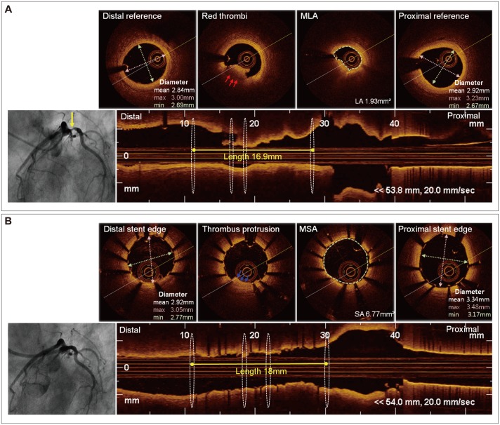
The significant morbidity and mortality associated with coronary artery disease has spurred the development of intravascular imaging devices to optimize the detection and assessment of coronary lesions and percutaneous coronary interventions. Intravascular ultrasound (IVUS) uses reflected ultrasound waves to quantitatively and qualitatively assess lesions; integrated backscatter and virtual histology IVUS more precisely characterizes plaque composition; angioscopy directly visualize thrombus and plaque; optical coherence tomography using near-infrared (NIR) light with very high spatial resolution provides more accurate images; and the recently introduced NIR spectroscopy identifies chemical components in coronary artery plaques based on differential light absorption in the NIR spectrum. This article reviews usefulness of these devices and hybrids thereof.

摘要

与冠状动脉疾病相关的显著发病率和死亡率促使了血管内成像设备的发展,以优化冠状动脉病变和经皮冠状动脉介入治疗的检测和评估。血管内超声(IVUS)使用反射超声波对病变进行定量和定性评估;背向散射积分和虚拟组织学 IVUS 更精确地描述斑块成分;血管内镜直接观察血栓和斑块;近红外光(NIR)的光学相干断层扫描具有非常高的空间分辨率,提供更准确的图像;以及最近推出的 NIR 光谱学根据 NIR 光谱中的差分光吸收来识别冠状动脉斑块中的化学成分。本文综述了这些设备及其杂交体的用途。

https://cdn.ncbi.nlm.nih.gov/pmc/blobs/580b/4372986/72d103518594/kcj-45-87-g002.jpg
https://cdn.ncbi.nlm.nih.gov/pmc/blobs/580b/4372986/e271457aa390/kcj-45-87-g001.jpg
https://cdn.ncbi.nlm.nih.gov/pmc/blobs/580b/4372986/72d103518594/kcj-45-87-g002.jpg
https://cdn.ncbi.nlm.nih.gov/pmc/blobs/580b/4372986/e271457aa390/kcj-45-87-g001.jpg
https://cdn.ncbi.nlm.nih.gov/pmc/blobs/580b/4372986/72d103518594/kcj-45-87-g002.jpg

相似文献

1
Practical application of coronary imaging devices in cardiovascular intervention.冠状动脉成像设备在心血管介入中的实际应用。
Korean Circ J. 2015 Mar;45(2):87-95. doi: 10.4070/kcj.2015.45.2.87. Epub 2015 Mar 24.
2
imaging of vulnerable plaque with intravascular modalities: its advantages and limitations.血管内成像技术对易损斑块的成像:优势与局限
Cardiovasc Diagn Ther. 2020 Oct;10(5):1461-1479. doi: 10.21037/cdt-20-238.
3
The Role of Intracoronary Plaque Imaging with Intravascular Ultrasound, Optical Coherence Tomography, and Near-Infrared Spectroscopy in Patients with Coronary Artery Disease.冠状动脉内斑块成像(血管内超声、光学相干断层扫描和近红外光谱)在冠状动脉疾病患者中的作用。
Curr Atheroscler Rep. 2016 Sep;18(9):57. doi: 10.1007/s11883-016-0607-0.
4
Current diagnostic modalities for vulnerable plaque detection.用于易损斑块检测的当前诊断方法。
Curr Pharm Des. 2007;13(10):995-1001. doi: 10.2174/138161207780487511.
5
Application of virtual histological intravascular ultrasound in plaque composition assessment of saphenous vein graft diseases.虚拟组织学血管内超声在评估大隐静脉桥病变斑块成分中的应用。
Chin Med J (Engl). 2019 Apr 20;132(8):957-962. doi: 10.1097/CM9.0000000000000183.
6
Invasive imaging of the coronary atherosclerotic plaque.冠状动脉粥样硬化斑块的侵入性成像。
Minerva Cardioangiol. 2012 Jun;60(3):305-29.
7
Intracoronary imaging modalities for vulnerable plaques.用于检测易损斑块的冠状动脉内成像技术
J Nippon Med Sch. 2011;78(6):340-51. doi: 10.1272/jnms.78.340.
8
Additive Value of Integrated Backscatter IVUS for Detection of Vulnerable Plaque by Optical Frequency Domain Imaging: An Ex Vivo Autopsy Study of Human Coronary Arteries.光学相干断层成像技术检测易损斑块的血管内超声背向散射积分的附加价值:人体冠状动脉尸检研究。
JACC Cardiovasc Imaging. 2016 Feb;9(2):163-72. doi: 10.1016/j.jcmg.2015.07.011. Epub 2016 Jan 6.
9
Near-Infrared Spectroscopy Intravascular Ultrasound Imaging: State of the Art.近红外光谱血管内超声成像:现状
Front Cardiovasc Med. 2020 Jun 30;7:107. doi: 10.3389/fcvm.2020.00107. eCollection 2020.
10
In vivo quantitative tissue characterization of human coronary arterial plaques by use of integrated backscatter intravascular ultrasound and comparison with angioscopic findings.利用背向散射积分血管内超声对人体冠状动脉斑块进行体内定量组织特征分析并与血管内镜检查结果比较。
Circulation. 2002 May 28;105(21):2487-92. doi: 10.1161/01.cir.0000017200.47342.10.

引用本文的文献

1
Formation and Transformation of Neointima after Drug-eluting Stent Implantation: Insights from Optical Coherence Tomographic Studies.药物洗脱支架植入术后新生内膜的形成与转变:光学相干断层扫描研究的见解
Korean Circ J. 2017 Nov;47(6):823-832. doi: 10.4070/kcj.2017.0157. Epub 2017 Oct 19.
2
Characteristics of Earlier Versus Delayed Presentation of Very Late Drug-Eluting Stent Thrombosis: An Optical Coherence Tomographic Study.极晚期药物洗脱支架血栓形成的早期与延迟表现特征:一项光学相干断层扫描研究
J Am Heart Assoc. 2017 Apr 14;6(4):e005386. doi: 10.1161/JAHA.116.005386.
3
Multimodality Intravascular Imaging Assessment of Plaque Erosion versus Plaque Rupture in Patients with Acute Coronary Syndrome.

本文引用的文献

1
Cutting-balloon angioplasty before drug-eluting stent implantation for the treatment of severely calcified coronary lesions.切割球囊血管成形术在药物洗脱支架植入术之前治疗严重钙化的冠状动脉病变。
J Geriatr Cardiol. 2014 Mar;11(1):44-9. doi: 10.3969/j.issn.1671-5411.2014.01.012.
2
Meta-analysis of outcomes after intravascular ultrasound-guided versus angiography-guided drug-eluting stent implantation in 26,503 patients enrolled in three randomized trials and 14 observational studies.26503 例患者的 3 项随机试验和 14 项观察性研究的血管内超声指导与血管造影指导药物洗脱支架植入术后结局的荟萃分析。
Am J Cardiol. 2014 Apr 15;113(8):1338-47. doi: 10.1016/j.amjcard.2013.12.043. Epub 2014 Jan 31.
3
急性冠状动脉综合征患者斑块侵蚀与斑块破裂的多模态血管内成像评估
Korean Circ J. 2016 Jul;46(4):499-506. doi: 10.4070/kcj.2016.46.4.499. Epub 2016 Jul 21.
Optical coherence tomography-guided primary percutaneous coronary intervention in ST-segment elevation myocardial infarction patients: a pilot study.
光学相干断层扫描引导下的 ST 段抬高型心肌梗死患者直接经皮冠状动脉介入治疗:一项初步研究。
Can J Cardiol. 2014 Apr;30(4):420-7. doi: 10.1016/j.cjca.2013.12.016. Epub 2013 Dec 30.
4
Optical coherence tomography guided PCI - initial experience at Apollo Health City, Jubilee Hills, Hyderabad.光学相干断层扫描引导下的经皮冠状动脉介入治疗——在海得拉巴朱比利山阿波罗健康城的初步经验。
Indian Heart J. 2014 Jan-Feb;66(1):31-7. doi: 10.1016/j.ihj.2013.12.048. Epub 2014 Jan 17.
5
Intravascular ultrasound-guided implantation of drug-eluting stents to improve outcome: a meta-analysis.血管内超声指导下药物洗脱支架置入术改善预后的Meta 分析。
JACC Cardiovasc Interv. 2014 Mar;7(3):233-43. doi: 10.1016/j.jcin.2013.09.013. Epub 2014 Feb 13.
6
Incidences, predictors, and clinical outcomes of acute and late stent malapposition detected by optical coherence tomography after drug-eluting stent implantation.光学相干断层成像术检测药物洗脱支架置入术后急性和晚期支架贴壁不良的发生率、预测因素及临床转归。
Circ Cardiovasc Interv. 2014 Feb;7(1):88-96. doi: 10.1161/CIRCINTERVENTIONS.113.000797. Epub 2014 Jan 14.
7
Relationship between intravascular ultrasound guidance and clinical outcomes after drug-eluting stents: the assessment of dual antiplatelet therapy with drug-eluting stents (ADAPT-DES) study.血管内超声指导与药物洗脱支架治疗后临床结局的关系:药物洗脱支架双重抗血小板治疗评估(ADAPT-DES)研究。
Circulation. 2014 Jan 28;129(4):463-70. doi: 10.1161/CIRCULATIONAHA.113.003942. Epub 2013 Nov 26.
8
Quantitative angiography and optical coherence tomography for the functional assessment of nonobstructive coronary stenoses: comparison with fractional flow reserve.定量血管造影和光相干断层成像术评估非阻塞性冠状动脉狭窄的功能:与血流储备分数的比较。
Am Heart J. 2013 Dec;166(6):1010-1018.e1. doi: 10.1016/j.ahj.2013.08.016. Epub 2013 Oct 17.
9
OCT compared with IVUS in a coronary lesion assessment: the OPUS-CLASS study.OCT 与 IVUS 在冠状动脉病变评估中的比较:OPUS-CLASS 研究。
JACC Cardiovasc Imaging. 2013 Oct;6(10):1095-1104. doi: 10.1016/j.jcmg.2013.04.014. Epub 2013 Sep 4.
10
Optical coherence tomography criteria for defining functional severity of intermediate lesions: a comparative study with FFR.光学相干断层成像标准定义中间病变的功能严重程度:与 FFR 的对比研究。
Int J Cardiovasc Imaging. 2013 Dec;29(8):1685-91. doi: 10.1007/s10554-013-0283-x. Epub 2013 Sep 3.