Arvanitakis Zoe, Fleischman Debra A, Arfanakis Konstantinos, Leurgans Sue E, Barnes Lisa L, Bennett David A
Rush Alzheimer's Disease Center, Rush University Medical Center, 600 S. Paulina Ave, Suite 1020, Chicago, IL, 60612, USA.
Department of Neurological Sciences, Rush University Medical Center, Chicago, USA.
Brain Struct Funct. 2016 May;221(4):2135-46. doi: 10.1007/s00429-015-1034-7. Epub 2015 Apr 2.
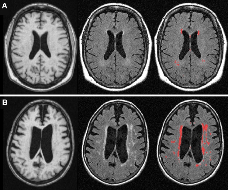
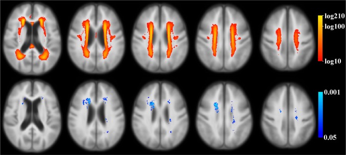
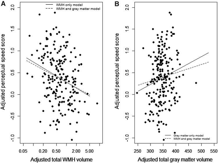
Both presence of white matter hyperintensities (WMH) and smaller total gray matter volume on brain magnetic resonance imaging (MRI) are common findings in old age, and contribute to impaired cognition. We tested whether total WMH volume and gray matter volume had independent associations with cognition in community-dwelling individuals without dementia or mild cognitive impairment (MCI). We used data from participants of the Rush Memory and Aging Project. Brain MRI was available in 209 subjects without dementia or MCI (mean age 80; education = 15 years; 74 % women). WMH and gray matter were automatically segmented, and the total WMH and gray matter volumes were measured. Both MRI-derived measures were normalized by the intracranial volume. Cognitive data included composite measures of five different cognitive domains, based on 19 individual tests. Linear regression analyses, adjusted for age, sex, and education, were used to examine the relationship of logarithmically-transformed total WMH volume and of total gray matter volume to cognition. Larger total WMH volumes were associated with lower levels of perceptual speed (p < 0.001), but not with episodic memory, semantic memory, working memory, or visuospatial abilities (all p > 0.10). Smaller total gray matter volumes were associated with lower levels of perceptual speed (p = 0.013) and episodic memory (p = 0.001), but not with the other three cognitive domains (all p > 0.14). Larger total WMH volume was correlated with smaller total gray matter volume (p < 0.001). In a model with both MRI-derived measures included, the relation of WMH to perceptual speed remained significant (p < 0.001), while gray matter volumes were no longer related (p = 0.14). This study of older community-dwelling individuals without overt cognitive impairment suggests that the association of larger total WMH volume with lower perceptual speed is independent of total gray matter volume. These results help elucidate the pathological processes leading to lower cognitive function in aging.
脑磁共振成像(MRI)显示的白质高信号(WMH)和脑灰质总体积减小在老年人中均为常见表现,并会导致认知功能受损。我们测试了在无痴呆或轻度认知障碍(MCI)的社区居住个体中,WMH总体积和灰质体积是否与认知功能存在独立关联。我们使用了拉什记忆与衰老项目参与者的数据。209名无痴呆或MCI的受试者(平均年龄80岁;受教育年限 = 15年;74%为女性)有脑MRI数据。WMH和灰质自动分割后测量其总体积。两种MRI衍生测量值均通过颅内体积进行标准化。认知数据包括基于19项个体测试的五个不同认知领域的综合测量值。采用经年龄、性别和受教育程度校正的线性回归分析,以检验对数转换后的WMH总体积和灰质总体积与认知功能的关系。WMH总体积越大,感知速度水平越低(p < 0.001),但与情景记忆、语义记忆、工作记忆或视觉空间能力无关(所有p > 0.10)。灰质总体积越小,感知速度水平越低(p = 0.013)和情景记忆越低(p = 0.001),但与其他三个认知领域无关(所有p > 0.14)。WMH总体积越大与灰质总体积越小相关(p < 0.001)。在同时纳入两种MRI衍生测量值的模型中,WMH与感知速度的关系仍然显著(p < 0.001),而灰质体积不再相关(p = 0.14)。这项针对无明显认知障碍的老年社区居住个体的研究表明,WMH总体积较大与较低感知速度之间的关联独立于灰质总体积。这些结果有助于阐明导致衰老过程中认知功能下降的病理过程。