Winnicka Katarzyna, Wroblewska Magdalena, Sosnowska Katarzyna, Car Halina, Kasacka Irena
Department of Pharmaceutical Technology, Faculty of Pharmacy, Medical University of Białystok, Białystok, Poland.
Department of Experimental Pharmacology, Faculty of Health Sciences, Medical University of Białystok, Białystok, Poland.
Drug Des Devel Ther. 2015 Mar 5;9:1367-77. doi: 10.2147/DDDT.S78336. eCollection 2015.
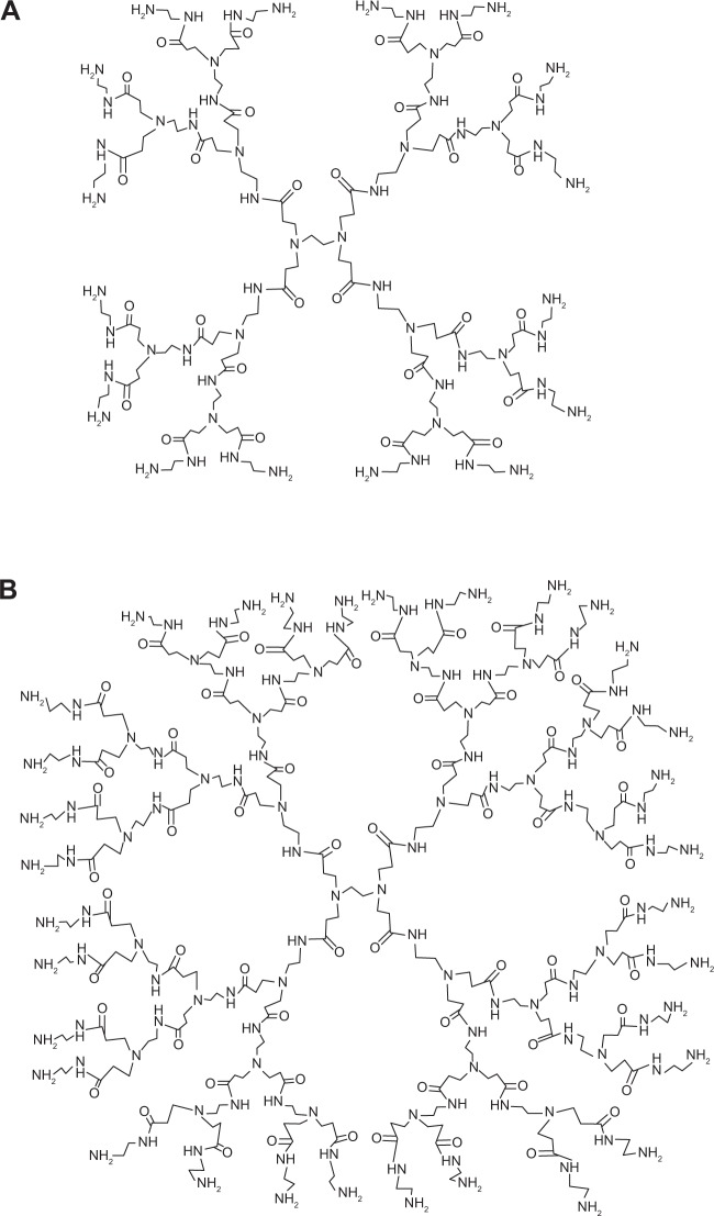
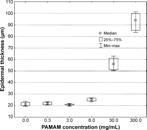
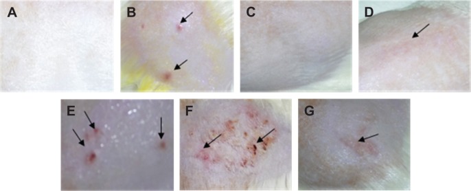
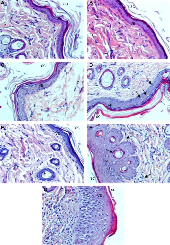
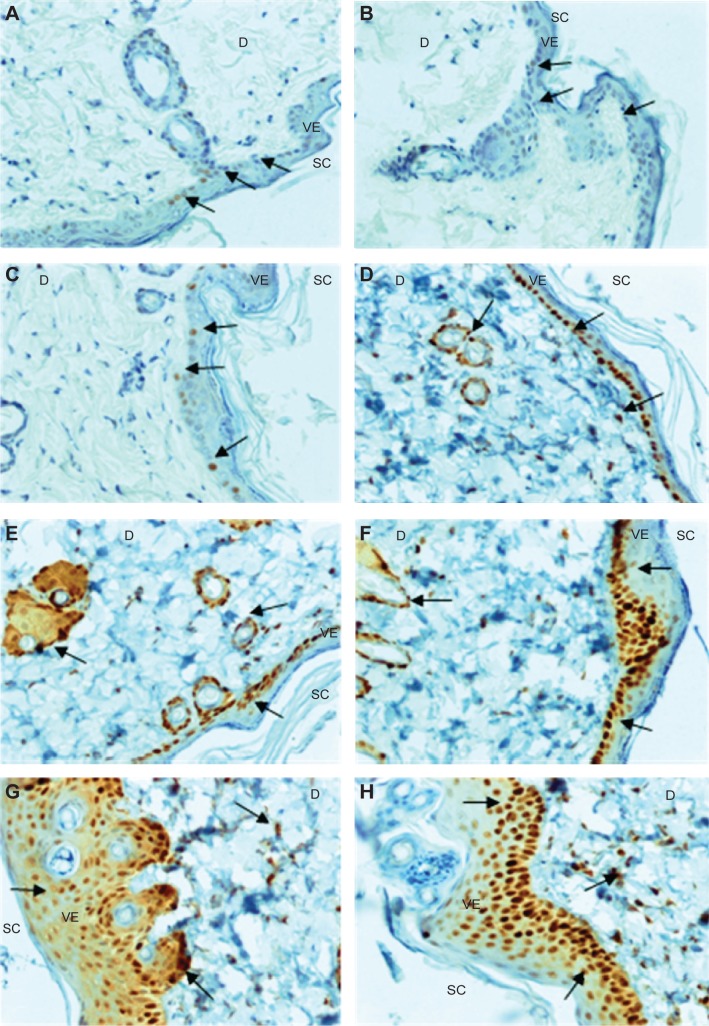
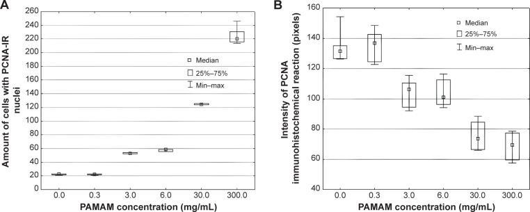
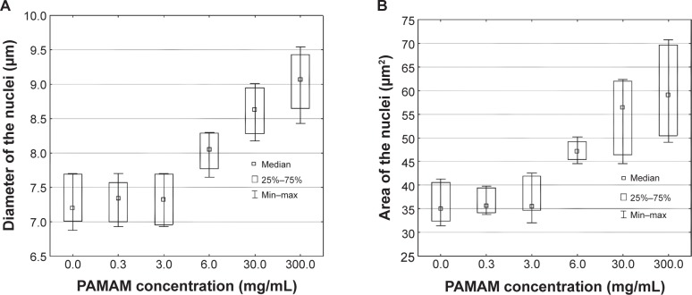
Polyamidoamine (PAMAM) dendrimers are multi-branched, three-dimensional polymers with unique architecture, which makes these molecules attractive for medical and pharmaceutical applications. Using PAMAM as drug carriers for topical delivery might be beneficial as they only produce a transient effect without skin irritation. To evaluate the dermal toxicity of cationic PAMAM dendrimers generation 2 and generation 3, skin irritation studies were performed in vivo in the rat skin model. After 10 days topical application of various concentrations of PAMAM-NH2 (0.3 mg/mL, 3 mg/mL, 6 mg/mL, 30 mg/mL, 300 mg/mL), skin irritation was evaluated by visual, histopathological, and immunohistochemical examination. Microscopic assessment after hematoxylin-eosin staining revealed significant morphological changes of epidermal cells after application of PAMAM-NH2 at a concentration of ≥6 mg/mL. Morphological alterations of epidermal cells included cytoplasmic vacuolization of keratinocytes in the basal and spinous layers. Cytomorphological changes in keratinocytes, overall picture of the epidermis, and histopathological changes in the dermis were dose dependent. Detected alterations concerned hyperplasia of connective tissue fibers and leukocyte infiltration. Visible granulocyte infiltration in the upper dermis and sockets formed by necrotic, cornified cells in the hyperplastic foci of epithelium were also noted. Immunohistochemical analyses revealed that increased nuclear immunoreactivity to PCNA correlated with the concentration of PAMAM-NH2, but no significant differences in the cell proliferation activity in skin treated with PAMAM-NH2 generation 2 or generation 3 were observed. Significantly higher expression of PCNA extended throughout the skin layers might suggest abnormal cell proliferation, which, as a consequence, might even lead to neoplastic changes.
聚酰胺 - 胺(PAMAM)树枝状大分子是具有独特结构的多分支三维聚合物,这使得这些分子在医学和制药应用中具有吸引力。将PAMAM用作局部给药的药物载体可能是有益的,因为它们只会产生短暂的效果而不会引起皮肤刺激。为了评估第2代和第3代阳离子PAMAM树枝状大分子的皮肤毒性,在大鼠皮肤模型中进行了体内皮肤刺激研究。在局部应用各种浓度的PAMAM - NH2(0.3 mg/mL、3 mg/mL、6 mg/mL、30 mg/mL、300 mg/mL)10天后,通过视觉、组织病理学和免疫组织化学检查评估皮肤刺激情况。苏木精 - 伊红染色后的显微镜评估显示,在应用浓度≥6 mg/mL的PAMAM - NH2后,表皮细胞出现了明显的形态学变化。表皮细胞的形态学改变包括基底层和棘层角质形成细胞的细胞质空泡化。角质形成细胞的细胞形态学变化、表皮的整体情况以及真皮的组织病理学变化均呈剂量依赖性。检测到的改变包括结缔组织纤维增生和白细胞浸润。在上层真皮中可见粒细胞浸润,并且在上皮增生灶中也观察到由坏死、角化细胞形成的窝。免疫组织化学分析显示,对PCNA的核免疫反应性增加与PAMAM - NH2的浓度相关,但在用第2代或第3代PAMAM - NH2处理的皮肤中,未观察到细胞增殖活性的显著差异。PCNA在整个皮肤层中显著更高的表达可能表明细胞增殖异常,这可能甚至导致肿瘤性变化。