Całkosiński Ireneusz, Dobrzyński Maciej, Rosińczuk Joanna, Dudek Krzysztof, Chrószcz Aleksander, Fita Katarzyna, Dymarek Robert
Department of Nervous System Diseases, The Faculty of Health Science, Wroclaw Medical University, 5 Bartla Street, 51-618 Wroclaw, Poland.
Department of Conservative Dentistry and Pedodontics, The Faculty of Dentistry, Wroclaw Medical University, 26 Krakowska Street, 50-425 Wroclaw, Poland.
Biomed Res Int. 2015;2015:972535. doi: 10.1155/2015/972535. Epub 2015 Mar 5.
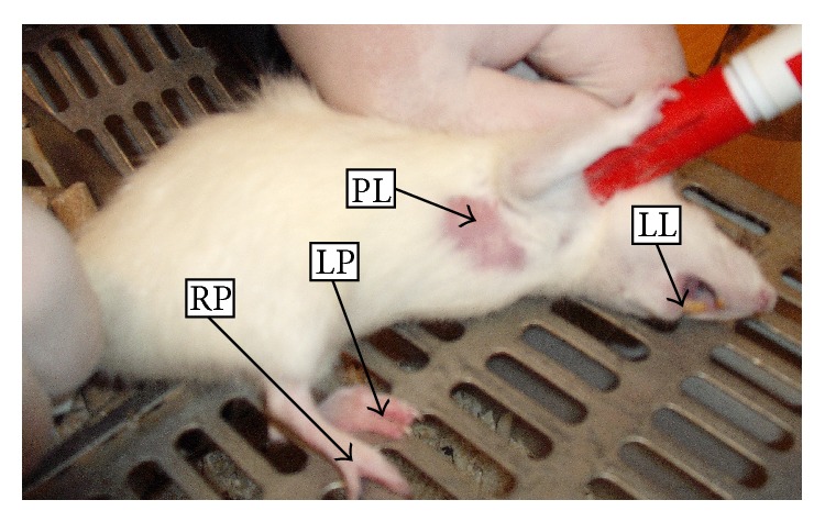
Thermographic assessment of temperature distribution within the examined tissues allows a quick, noncontact, noninvasive measurement of their temperature. The aim of the study was to evaluate the usefulness of digital infrared imaging in monitoring experimental inflammation of pleura (PL), lower lip (LL), and left paw (LP) and right paw (RP) of lower limbs in rats.
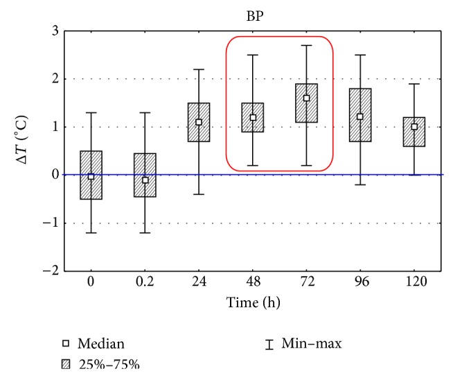
The inflammatory reaction was induced by injection of 1% carrageenin solution into pleural cavity, lip, or paws. With the use of digital infrared imaging temperature measurement was conducted at 0 to 72 hours of the inflammatory reaction.
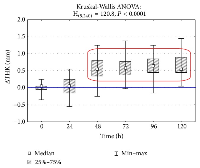
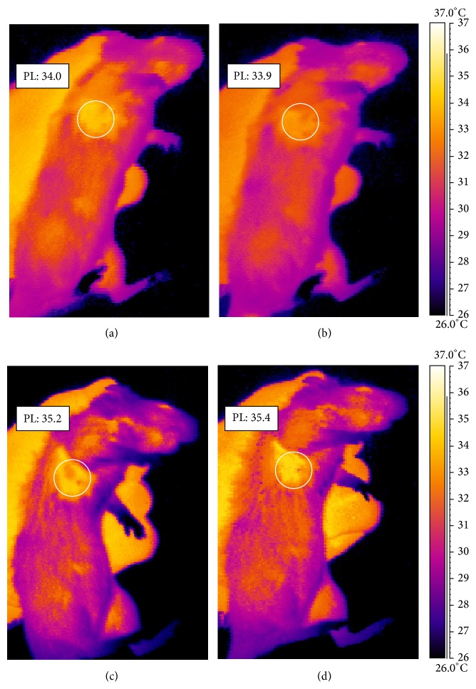
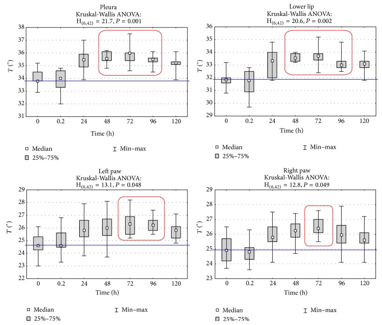
The temperature decrease was observed at the site of injection directly afterwards. Next, it was gradually increasing and it reached the maximum on the third day of the inflammatory reaction. Statistically significant changes were observed after 48-hour period in PL and LL regions, as well as after 72-hour period in LP and RP regions (P < 0.005).
It was found that thermographic examination allows for indicating the presence of inflammatory reaction within examined tissues and determining the dynamics of this process. This method could be used as alternative procedure that allows using fewer animals for experiments.
通过热成像评估被检查组织内的温度分布,可实现对其温度的快速、非接触、无创测量。本研究旨在评估数字红外成像在监测大鼠胸膜(PL)、下唇(LL)以及下肢左爪(LP)和右爪(RP)的实验性炎症中的实用性。
通过向胸腔、唇部或爪子注射1%角叉菜胶溶液诱导炎症反应。利用数字红外成像在炎症反应的0至72小时进行温度测量。
注射后即刻在注射部位观察到温度下降。接下来,温度逐渐升高,并在炎症反应的第三天达到最高值。在PL和LL区域,48小时后观察到具有统计学意义的变化;在LP和RP区域,72小时后观察到具有统计学意义的变化(P < 0.005)。
发现热成像检查能够指示被检查组织内炎症反应的存在,并确定该过程的动态变化。此方法可作为一种替代程序,允许在实验中使用更少的动物。