• 文献检索
  • 文档翻译
  • 深度研究
  • 学术资讯
  • Suppr Zotero 插件Zotero 插件
  • 邀请有礼
  • 套餐&价格
  • 历史记录
应用&插件
Suppr Zotero 插件Zotero 插件浏览器插件Mac 客户端Windows 客户端微信小程序
定价
高级版会员购买积分包购买API积分包
服务
文献检索文档翻译深度研究API 文档MCP 服务
关于我们
关于 Suppr公司介绍联系我们用户协议隐私条款
关注我们

Suppr 超能文献

核心技术专利:CN118964589B侵权必究
粤ICP备2023148730 号-1Suppr @ 2026

文献检索

告别复杂PubMed语法,用中文像聊天一样搜索,搜遍4000万医学文献。AI智能推荐,让科研检索更轻松。

立即免费搜索

文件翻译

保留排版,准确专业,支持PDF/Word/PPT等文件格式,支持 12+语言互译。

免费翻译文档

深度研究

AI帮你快速写综述,25分钟生成高质量综述,智能提取关键信息,辅助科研写作。

立即免费体验

通过血管采样、血流速度分布及血管直径来表征人球结膜微血管系统

Vessel Sampling and Blood Flow Velocity Distribution With Vessel Diameter for Characterizing the Human Bulbar Conjunctival Microvasculature.

作者信息

Wang Liang, Yuan Jin, Jiang Hong, Yan Wentao, Cintrón-Colón Hector R, Perez Victor L, DeBuc Delia C, Feuer William J, Wang Jianhua

机构信息

Palmer Trinity School (L.W.), Miami, FL; Bascom Palmer Eye Institute (J.Y., H.J., H.R.C.-C., V.L.P., D.C.D., W.J.F., J.W.), Department of Ophthalmology, University of Miami, Miami, FL; Zhongshan Ophthalmic Centre (J.Y.), Sun Yat-sen University, Department of Ophthalmology and Optometry, Guangzhou, China; School of Ophthalmology and Optometry (W.Y.), Wenzhou Medical College, Wenzhou, China; Departments of Neurology (H.J.), and Microbiology and Immunology (H.R.C.-C., V.L.P.), University of Miami, FL; and Statistic Division (W.J.F.), Bascom Palmer Eye Institute, University of Miami, Miami, FL.

出版信息

Eye Contact Lens. 2016 Mar;42(2):135-40. doi: 10.1097/ICL.0000000000000146.

DOI:10.1097/ICL.0000000000000146
PMID:25839347
原文链接:https://pmc.ncbi.nlm.nih.gov/articles/PMC4591084/
Abstract

PURPOSE

This study determined (1) how many vessels (i.e., the vessel sampling) are needed to reliably characterize the bulbar conjunctival microvasculature and (2) if characteristic information can be obtained from the distribution histogram of the blood flow velocity and vessel diameter.

METHODS

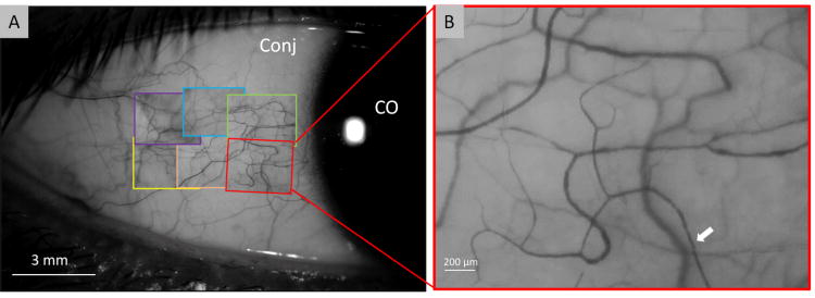
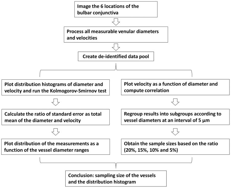
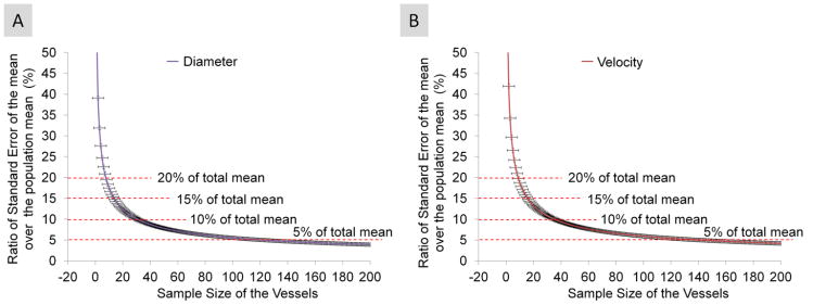
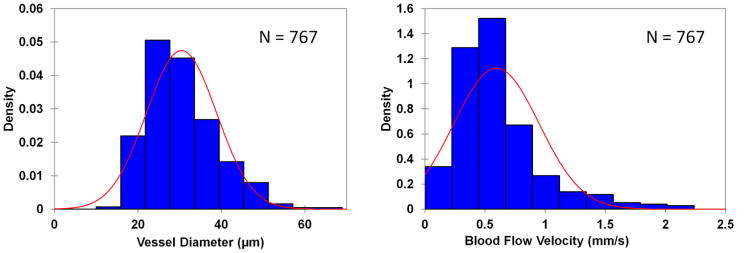
Functional slitlamp biomicroscope was used to image hundreds of venules per subject. The bulbar conjunctiva in five healthy human subjects was imaged on six different locations in the temporal bulbar conjunctiva. The histograms of the diameter and velocity were plotted to examine whether the distribution was normal. Standard errors were calculated from the standard deviation and vessel sample size. The ratio of the standard error of the mean over the population mean was used to determine the sample size cutoff. The velocity was plotted as a function of the vessel diameter to display the distribution of the diameter and velocity.

RESULTS

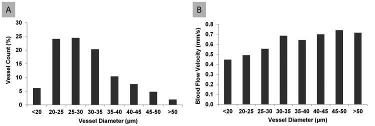
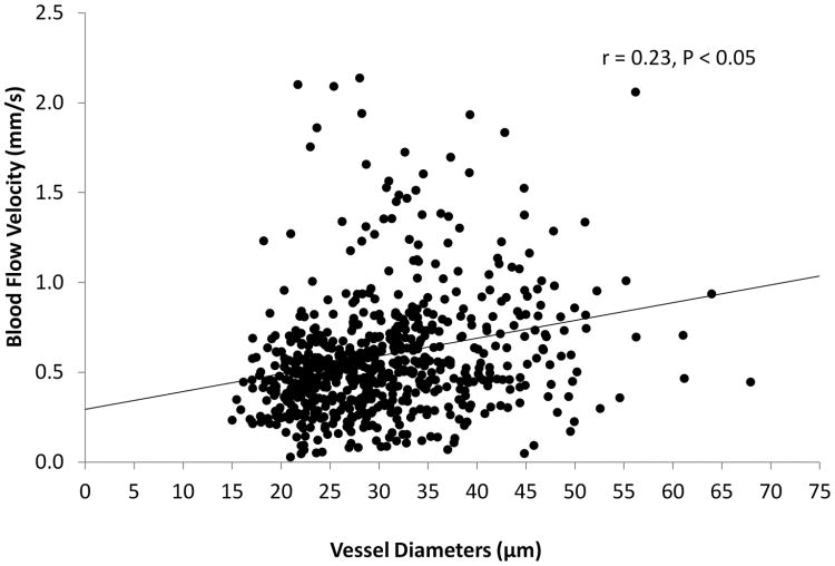
The results showed that the sampling size was approximately 15 vessels, which generated a standard error equivalent to 15% of the population mean from the total vessel population. The distributions of the diameter and velocity were not only unimodal, but also somewhat positively skewed and not normal. The blood flow velocity was related to the vessel diameter (r=0.23, P<0.05).

CONCLUSIONS

This was the first study to determine the sampling size of the vessels and the distribution histogram of the blood flow velocity and vessel diameter, which may lead to a better understanding of the human microvascular system of the bulbar conjunctiva.

摘要

目的

本研究确定了(1)可靠地表征球结膜微血管系统需要多少血管(即血管采样),以及(2)是否可以从血流速度和血管直径的分布直方图中获得特征信息。

方法

使用功能性裂隙灯生物显微镜对每位受试者的数百条小静脉进行成像。对5名健康人类受试者颞侧球结膜的6个不同位置进行球结膜成像。绘制直径和速度的直方图以检查分布是否呈正态分布。根据标准差和血管样本量计算标准误差。使用样本均值的标准误差与总体均值的比率来确定样本量截止值。绘制速度与血管直径的函数关系图以显示直径和速度的分布。

结果

结果表明,采样大小约为15条血管,其产生的标准误差相当于总血管群体中总体均值的15%。直径和速度的分布不仅是单峰的,而且有些正偏态且不呈正态分布。血流速度与血管直径相关(r = 0.23,P < 0.05)。

结论

这是第一项确定血管采样大小以及血流速度和血管直径分布直方图的研究,这可能有助于更好地理解球结膜的人体微血管系统。

https://cdn.ncbi.nlm.nih.gov/pmc/blobs/f6f9/4591084/a27bac53c2a6/nihms657402f6.jpg
https://cdn.ncbi.nlm.nih.gov/pmc/blobs/f6f9/4591084/f1ea823e8763/nihms657402f1.jpg
https://cdn.ncbi.nlm.nih.gov/pmc/blobs/f6f9/4591084/ee3d4bf77669/nihms657402f2.jpg
https://cdn.ncbi.nlm.nih.gov/pmc/blobs/f6f9/4591084/8ce47c8e5b42/nihms657402f3.jpg
https://cdn.ncbi.nlm.nih.gov/pmc/blobs/f6f9/4591084/f039fec3cb61/nihms657402f4.jpg
https://cdn.ncbi.nlm.nih.gov/pmc/blobs/f6f9/4591084/76c3f61c59cf/nihms657402f5.jpg
https://cdn.ncbi.nlm.nih.gov/pmc/blobs/f6f9/4591084/a27bac53c2a6/nihms657402f6.jpg

相似文献

1
Vessel Sampling and Blood Flow Velocity Distribution With Vessel Diameter for Characterizing the Human Bulbar Conjunctival Microvasculature.通过血管采样、血流速度分布及血管直径来表征人球结膜微血管系统
Eye Contact Lens. 2016 Mar;42(2):135-40. doi: 10.1097/ICL.0000000000000146.
2
Altered Bulbar Conjunctival Microcirculation in Response to Contact Lens Wear.因佩戴隐形眼镜而改变的球结膜微循环。
Eye Contact Lens. 2017 Mar;43(2):95-99. doi: 10.1097/ICL.0000000000000241.
3
Measurement variability of the bulbar conjunctival microvasculature in healthy subjects using functional slit lamp biomicroscopy (FSLB).使用功能性裂隙灯生物显微镜检查(FSLB)对健康受试者球结膜微血管系统的测量变异性。
Microvasc Res. 2015 Sep;101:15-9. doi: 10.1016/j.mvr.2015.05.003. Epub 2015 Jun 16.
4
Microvascular abnormalities in dry eye patients.干眼患者的微血管异常。
Microvasc Res. 2018 Jul;118:155-161. doi: 10.1016/j.mvr.2018.03.015. Epub 2018 Mar 28.
5
Quantitative assessment of conjunctival microvascular circulation of the human eye.定量评估人眼结膜微血管循环。
Microvasc Res. 2010 Mar;79(2):109-13. doi: 10.1016/j.mvr.2009.12.003. Epub 2010 Jan 4.
6
Functional slit lamp biomicroscopy for imaging bulbar conjunctival microvasculature in contact lens wearers.用于对隐形眼镜佩戴者球结膜微血管系统进行成像的功能性裂隙灯生物显微镜检查。
Microvasc Res. 2014 Mar;92:62-71. doi: 10.1016/j.mvr.2014.01.005. Epub 2014 Jan 17.
7
Quantitative assessment of the conjunctival microcirculation using a smartphone and slit-lamp biomicroscope.使用智能手机和裂隙灯生物显微镜对结膜微循环进行定量评估。
Microvasc Res. 2019 Nov;126:103907. doi: 10.1016/j.mvr.2019.103907. Epub 2019 Jul 19.
8
Bulbar Conjunctival Microvascular Alterations in Thyroid-Associated Ophthalmopathy Patients with Different Activities.甲状腺相关眼病不同活动度患者球结膜微血管改变。
Curr Eye Res. 2021 Jul;46(7):943-948. doi: 10.1080/02713683.2020.1849729. Epub 2021 Mar 15.
9
Changes in bulbar conjunctival microcirculation and microvasculature during short-term scleral lens wearing and their associated factors.短期巩膜镜佩戴过程中球结膜微循环和微血管的变化及其相关因素。
Cont Lens Anterior Eye. 2024 Oct;47(5):102159. doi: 10.1016/j.clae.2024.102159. Epub 2024 Jun 8.
10
In vivo oximetry of human bulbar conjunctival and episcleral microvasculature using snapshot multispectral imaging.使用快照多光谱成像技术对人球结膜和巩膜上微血管进行体内血氧测定。
Exp Eye Res. 2016 Aug;149:48-58. doi: 10.1016/j.exer.2016.06.008. Epub 2016 Jun 15.

引用本文的文献

1
Identification and quantification of micro-nano-plastics in polypropylene-bottled injections.聚丙烯瓶装注射液中微纳米塑料的鉴定与定量分析
Heliyon. 2024 Jul 27;10(15):e35101. doi: 10.1016/j.heliyon.2024.e35101. eCollection 2024 Aug 15.
2
Computational analysis of cancer cell adhesion in curved vessels affected by wall shear stress for prediction of metastatic spreading.受壁面剪应力影响的弯曲血管中癌细胞黏附的计算分析,用于预测转移扩散。
Front Bioeng Biotechnol. 2024 May 27;12:1393413. doi: 10.3389/fbioe.2024.1393413. eCollection 2024.
3
Development of a perfusable, hierarchical microvasculature-on-a-chip model.

本文引用的文献

1
Functional slit lamp biomicroscopy for imaging bulbar conjunctival microvasculature in contact lens wearers.用于对隐形眼镜佩戴者球结膜微血管系统进行成像的功能性裂隙灯生物显微镜检查。
Microvasc Res. 2014 Mar;92:62-71. doi: 10.1016/j.mvr.2014.01.005. Epub 2014 Jan 17.
2
Automated grading system for evaluation of ocular redness associated with dry eye.用于评估与干眼相关的眼部发红的自动分级系统。
Clin Ophthalmol. 2013;7:1197-1204. doi: 10.2147/OPTH.S39703. Epub 2013 Jun 20.
3
Human bulbar conjunctival hemodynamics in hemoglobin SS and SC disease.
可灌注的层次化微脉管芯片模型的开发。
Lab Chip. 2023 Oct 10;23(20):4552-4564. doi: 10.1039/d3lc00512g.
4
The potential role of ischaemia-reperfusion injury in chronic, relapsing diseases such as rheumatoid arthritis, Long COVID, and ME/CFS: evidence, mechanisms, and therapeutic implications.缺血再灌注损伤在类风湿性关节炎、长新冠和 ME/CFS 等慢性、复发性疾病中的潜在作用:证据、机制和治疗意义。
Biochem J. 2022 Aug 31;479(16):1653-1708. doi: 10.1042/BCJ20220154.
5
Identifying diabetes from conjunctival images using a novel hierarchical multi-task network.使用新型分层多任务网络从结膜图像中识别糖尿病。
Sci Rep. 2022 Jan 7;12(1):264. doi: 10.1038/s41598-021-04006-z.
6
Microfluidic model of monocyte extravasation reveals the role of hemodynamics and subendothelial matrix mechanics in regulating endothelial integrity.单核细胞渗出的微流控模型揭示了血流动力学和内皮下基质力学在调节内皮完整性中的作用。
Biomicrofluidics. 2021 Sep 14;15(5):054102. doi: 10.1063/5.0061997. eCollection 2021 Sep.
7
Quantification of Blood Flow Velocity in the Human Conjunctival Microvessels Using Deep Learning-Based Stabilization Algorithm.利用基于深度学习的稳定化算法对人结膜微血管血流速度进行定量分析。
Sensors (Basel). 2021 May 6;21(9):3224. doi: 10.3390/s21093224.
8
Assessment of the conjunctival microcirculation for patients presenting with acute myocardial infarction compared to healthy controls.评估急性心肌梗死患者与健康对照者的结膜微循环。
Sci Rep. 2021 Apr 7;11(1):7660. doi: 10.1038/s41598-021-87315-7.
9
Improved conjunctival microcirculation in diabetic retinopathy patients with MTHFR polymorphisms after Ocufolin™ Administration.奥可明®给药后,伴有 MTHFR 多态性的糖尿病视网膜病变患者的眼结合膜微循环得到改善。
Microvasc Res. 2020 Nov;132:104066. doi: 10.1016/j.mvr.2020.104066. Epub 2020 Aug 27.
10
A review of functional slit lamp biomicroscopy.功能性裂隙灯生物显微镜检查综述。
Eye Vis (Lond). 2019 May 21;6:15. doi: 10.1186/s40662-019-0140-7. eCollection 2019.
血红蛋白 SS 和 SC 病患者的人球结膜血液动力学。
Am J Hematol. 2013 Aug;88(8):661-4. doi: 10.1002/ajh.23475. Epub 2013 Jun 12.
4
Wall shear stress quantification in the human conjunctival pre-capillary arterioles in vivo.活体人结膜毛细血管前小动脉壁切应力定量分析。
Microvasc Res. 2013 Jan;85:34-9. doi: 10.1016/j.mvr.2012.11.003. Epub 2012 Nov 12.
5
Exchange transfusion therapy and its effects on real-time microcirculation in pediatric sickle cell anemia patients: an intravital microscopy study.换血疗法及其对小儿镰状细胞贫血患者实时微循环的影响:一项活体显微镜研究。
J Pediatr Hematol Oncol. 2012 Apr;34(3):169-74. doi: 10.1097/MPH.0b013e31823c27ef.
6
Microvascular abnormalities in the bulbar conjunctiva of contact lens users.接触镜使用者球结膜微血管异常。
Clin Hemorheol Microcirc. 2012;51(1):77-86. doi: 10.3233/CH-2011-1513.
7
Comparison of real-time microvascular abnormalities in pediatric and adult sickle cell anemia patients.比较儿童和成年镰状细胞贫血患者实时微血管异常。
Am J Hematol. 2010 Nov;85(11):899-901. doi: 10.1002/ajh.21853.
8
Alzheimer's disease: more than amyloid.阿尔茨海默病:不只是淀粉样蛋白。
Clin Hemorheol Microcirc. 2010;46(1):69-73. doi: 10.3233/CH-2010-1356.
9
Blood velocity pulse quantification in the human conjunctival pre-capillary arterioles.人结膜毛细血管前小动脉血流速度脉冲定量。
Microvasc Res. 2010 Sep;80(2):202-8. doi: 10.1016/j.mvr.2010.05.001. Epub 2010 May 18.
10
Quantitative assessment of conjunctival microvascular circulation of the human eye.定量评估人眼结膜微血管循环。
Microvasc Res. 2010 Mar;79(2):109-13. doi: 10.1016/j.mvr.2009.12.003. Epub 2010 Jan 4.