Motl Robert W, Pilutti Lara A, Hubbard Elizabeth A, Wetter Nathan C, Sosnoff Jacob J, Sutton Bradley P
Department of Kinesiology and Community Health, University of Illinois at Urbana-Champaign, IL, USA.
Department of Bioengineering, University of Illinois at Urbana-Champaign, IL, USA.
Neuroimage Clin. 2015 Feb 26;7:661-6. doi: 10.1016/j.nicl.2015.02.017. eCollection 2015.
There is little known about cardiorespiratory fitness and its association with volumes of the thalamus, hippocampus, and basal ganglia in multiple sclerosis (MS). Such inquiry is important for identifying a possible behavioral approach (e.g., aerobic exercise training) that might change volumes of deep gray matter (DGM) structures associated with cognitive and motor functions in MS.
This study examined the association between cardiorespiratory fitness and volumes of the thalamus, hippocampus, and basal ganglia in MS.
We enrolled 35 persons with MS who underwent a maximal exercise test for measuring cardiorespiratory fitness as peak oxygen consumption (VO2peak) and brain MRI. Volumes of the thalamus, hippocampus, caudate, putamen, and pallidum were calculated from 3D T1-weighted structural brain images. We examined associations using partial (pr) correlations controlling for demographic and clinical variables.
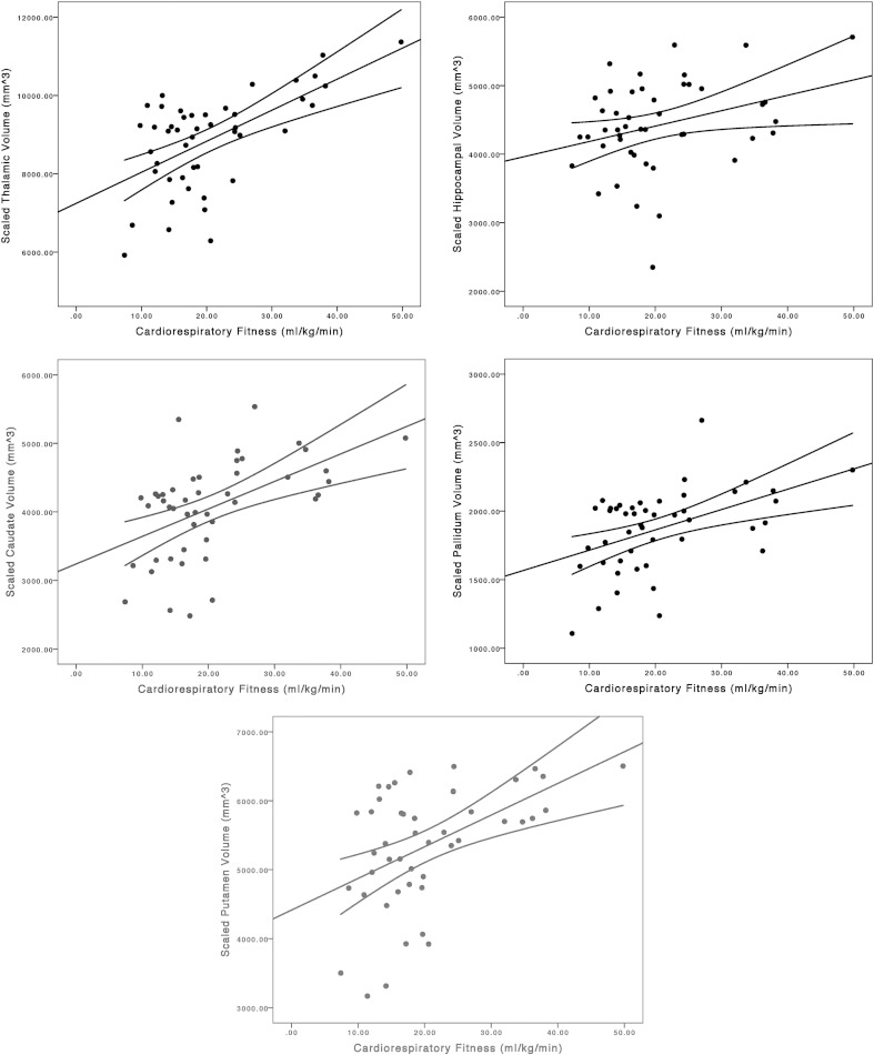
VO2peak was significantly associated with composite scaled volumes of the caudate(pr = .47, p < .01), putamen (pr = .44, p < .05), pallidum (pr = .40, p < .05), and hippocampus (pr = .42, p < .05), but not thalamus (pr = .31, p = .09), when controlling for sex, age, disability, and duration of MS.
Our results provide novel evidence that cardiorespiratory fitness is associated with volumes of DGM structures that are involved in motor and cognitive functions in MS.
关于多发性硬化症(MS)患者的心肺适能及其与丘脑、海马体和基底神经节体积的关联,目前所知甚少。此类研究对于确定一种可能改变与MS认知和运动功能相关的深部灰质(DGM)结构体积的行为方法(如有氧运动训练)具有重要意义。
本研究探讨了MS患者的心肺适能与丘脑、海马体和基底神经节体积之间的关联。
我们招募了35名MS患者,他们接受了最大运动测试以测量心肺适能,即峰值耗氧量(VO2peak),并进行了脑部MRI检查。从3D T1加权脑部结构图像中计算出丘脑、海马体、尾状核、壳核和苍白球的体积。我们使用控制人口统计学和临床变量的偏相关(pr)分析来研究关联。
在控制性别、年龄、残疾程度和MS病程后,VO2peak与尾状核的复合缩放体积(pr = 0.47,p < 0.01)、壳核(pr = 0.44,p < 0.05)、苍白球(pr = 0.40,p < 0.05)和海马体(pr = 0.42,p < 0.05)显著相关,但与丘脑(pr = 0.31,p = 0.09)无关。
我们的结果提供了新的证据,表明心肺适能与MS中参与运动和认知功能的DGM结构体积相关。