• 文献检索
  • 文档翻译
  • 深度研究
  • 学术资讯
  • Suppr Zotero 插件Zotero 插件
  • 邀请有礼
  • 套餐&价格
  • 历史记录
应用&插件
Suppr Zotero 插件Zotero 插件浏览器插件Mac 客户端Windows 客户端微信小程序
定价
高级版会员购买积分包购买API积分包
服务
文献检索文档翻译深度研究API 文档MCP 服务
关于我们
关于 Suppr公司介绍联系我们用户协议隐私条款
关注我们

Suppr 超能文献

核心技术专利:CN118964589B侵权必究
粤ICP备2023148730 号-1Suppr @ 2026

文献检索

告别复杂PubMed语法,用中文像聊天一样搜索,搜遍4000万医学文献。AI智能推荐,让科研检索更轻松。

立即免费搜索

文件翻译

保留排版,准确专业,支持PDF/Word/PPT等文件格式,支持 12+语言互译。

免费翻译文档

深度研究

AI帮你快速写综述,25分钟生成高质量综述,智能提取关键信息,辅助科研写作。

立即免费体验

表面涂层和 FePt 纳米粒子成分对抑制神经胶质瘤细胞增殖的影响。

Influences of surface coatings and components of FePt nanoparticles on the suppression of glioma cell proliferation.

机构信息

State Key Laboratory of Advanced Technology for Materials Synthesis and Processing and Biomedical Materials and Engineering Center, Wuhan University of Technology, Wuhan, China.

出版信息

Int J Nanomedicine. 2012;7:3295-307. doi: 10.2147/IJN.S32678. Epub 2012 Jul 6.

DOI:10.2147/IJN.S32678
PMID:22848161
原文链接:https://pmc.ncbi.nlm.nih.gov/articles/PMC3405879/
Abstract

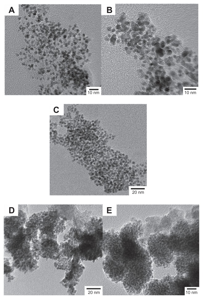
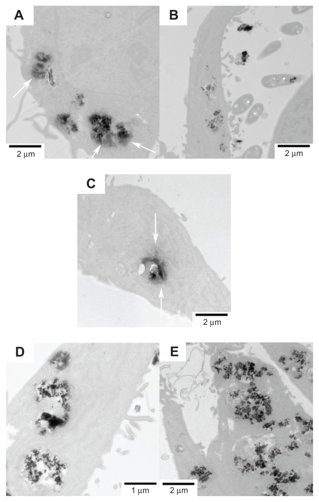
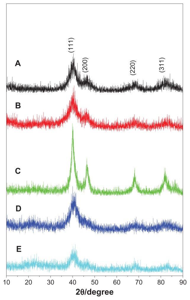
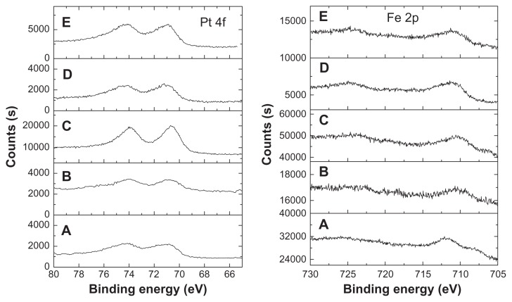
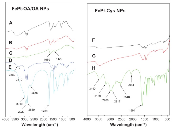
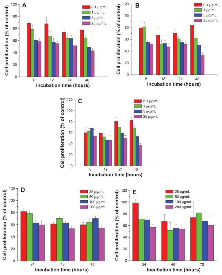
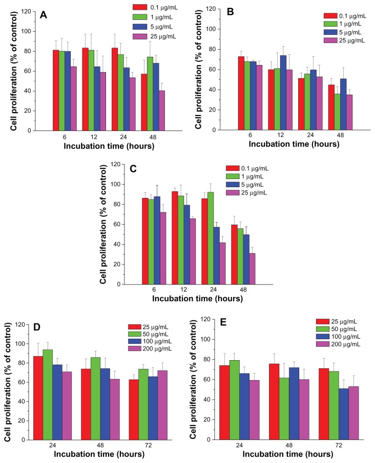
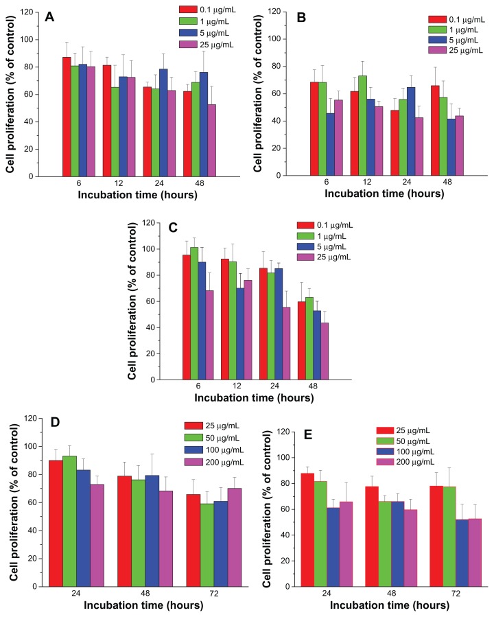
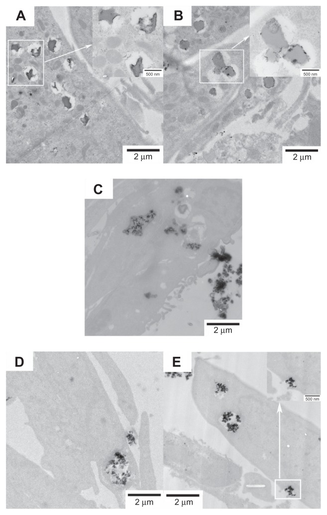
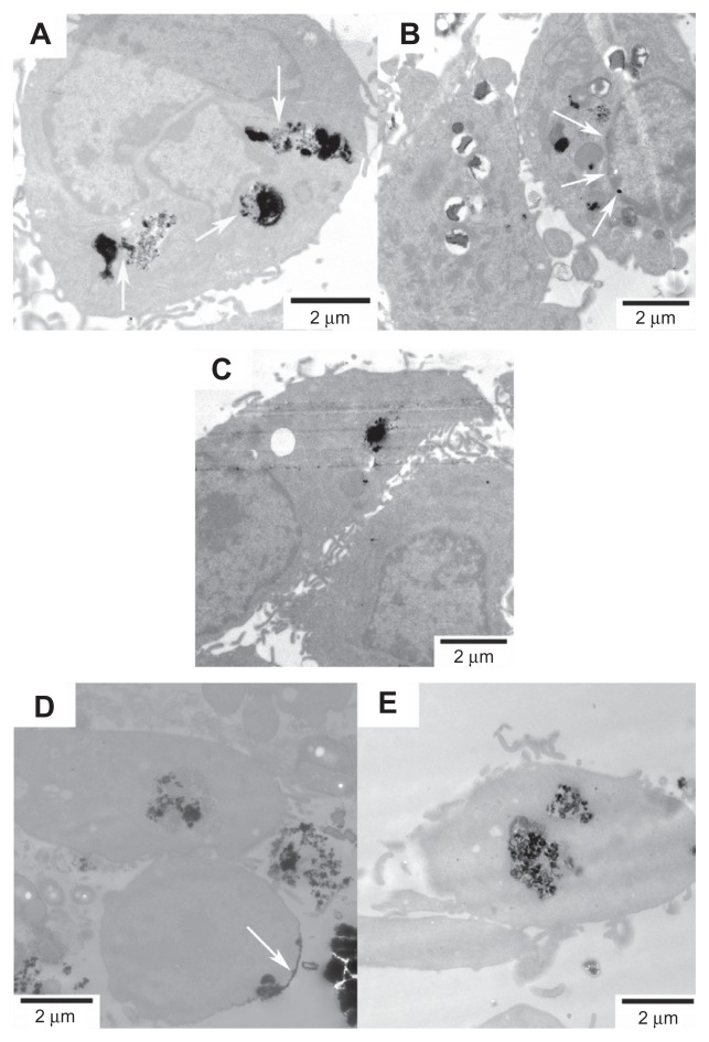
Malignant gliomas are primary brain tumors with high rates of morbidity and mortality; they are the fourth most common cause of cancer death. Novel diagnostic and therapeutic techniques based on nanomaterials provide promising options in the treatment of malignant gliomas. In order to evaluate the potential of FePt nanoparticles (NPs) for malignant glioma therapy, FePt NPs with different surface coatings and components were tunably synthesized using oleic acid/oleylamine (OA/OA) and cysteines (Cys) as the capping agents, respectively. The samples were characterized using X-ray diffraction, transmission electron microscopy (TEM), X-ray photon spectroscopy, Fourier transform infrared spectroscopy, atomic absorption spectrum, and zeta potential. The influence of the surface coatings and components of the FePt NPs on the proliferation of glioma cells was assessed through MTT assay and TEM observation using three typical glioma cell lines (glioma U251 cells, astrocytoma U87 cells, and neuroglioma H4 cells) as in vitro models. The results showed that the proliferation of glioma cells was significantly suppressed by lipophilic FePt-OA/OA NPs in a time- and/or dose-dependent manner, while no or low cytotoxic effects were detected in the case of hydrophilic FePt-Cys NPs. The IC₅₀ value of FePt-OA/OA NPs on the three glioma cell lines was approximately 5-10 μg mL⁻¹ after 24 hours' incubation. Although the cellular uptake of FePt NPs was confirmed regardless of the surface coatings and components of the FePt NPs, the suppression of FePt NPs on glioma cell proliferation was dominantly determined by their surface coatings rather than their components. Therefore, these results demonstrate that, through engineering of the surface coating, FePt NPs can potentially be developed as novel therapeutic agents for malignant gliomas.

摘要

恶性脑胶质瘤是一种高发病率和死亡率的原发性脑肿瘤;它是癌症死亡的第四大常见原因。基于纳米材料的新型诊断和治疗技术为恶性脑胶质瘤的治疗提供了有前景的选择。为了评估 FePt 纳米颗粒(NPs)在恶性脑胶质瘤治疗中的潜力,使用油酸/油胺(OA/OA)和半胱氨酸(Cys)分别作为封端剂,可调节合成具有不同表面涂层和组成的 FePt NPs。使用 X 射线衍射、透射电子显微镜(TEM)、X 射线光电子能谱、傅里叶变换红外光谱、原子吸收光谱和 Zeta 电位对样品进行了表征。通过 MTT 测定和使用三种典型的脑胶质瘤细胞系(脑胶质瘤 U251 细胞、星形细胞瘤 U87 细胞和神经胶质瘤 H4 细胞)作为体外模型的 TEM 观察,评估了 FePt NPs 的表面涂层和组成对神经胶质瘤细胞增殖的影响。结果表明,疏水性 FePt-OA/OA NPs 以时间和/或剂量依赖的方式显著抑制神经胶质瘤细胞的增殖,而亲水性 FePt-Cys NPs 则未检测到或低细胞毒性作用。在 24 小时孵育后,FePt-OA/OA NPs 对三种神经胶质瘤细胞系的 IC₅₀ 值约为 5-10μgmL⁻¹。尽管无论 FePt NPs 的表面涂层和组成如何,都确认了 FePt NPs 的细胞摄取,但抑制神经胶质瘤细胞增殖的主要因素是其表面涂层而不是其组成。因此,这些结果表明,通过表面涂层工程,FePt NPs 有可能被开发为恶性脑胶质瘤的新型治疗剂。

https://cdn.ncbi.nlm.nih.gov/pmc/blobs/acbe/3405879/5a1297353af3/ijn-7-3295f10.jpg
https://cdn.ncbi.nlm.nih.gov/pmc/blobs/acbe/3405879/524a37754203/ijn-7-3295f1.jpg
https://cdn.ncbi.nlm.nih.gov/pmc/blobs/acbe/3405879/617aea8db66b/ijn-7-3295f2.jpg
https://cdn.ncbi.nlm.nih.gov/pmc/blobs/acbe/3405879/7efc05c7f79b/ijn-7-3295f3.jpg
https://cdn.ncbi.nlm.nih.gov/pmc/blobs/acbe/3405879/b56d7f010dba/ijn-7-3295f4.jpg
https://cdn.ncbi.nlm.nih.gov/pmc/blobs/acbe/3405879/6087ec6dfac4/ijn-7-3295f5.jpg
https://cdn.ncbi.nlm.nih.gov/pmc/blobs/acbe/3405879/28a9466c7d01/ijn-7-3295f6.jpg
https://cdn.ncbi.nlm.nih.gov/pmc/blobs/acbe/3405879/101c110798a9/ijn-7-3295f7.jpg
https://cdn.ncbi.nlm.nih.gov/pmc/blobs/acbe/3405879/ff31c3f9acb9/ijn-7-3295f8.jpg
https://cdn.ncbi.nlm.nih.gov/pmc/blobs/acbe/3405879/0220ee7a737d/ijn-7-3295f9.jpg
https://cdn.ncbi.nlm.nih.gov/pmc/blobs/acbe/3405879/5a1297353af3/ijn-7-3295f10.jpg

相似文献

1
Influences of surface coatings and components of FePt nanoparticles on the suppression of glioma cell proliferation.表面涂层和 FePt 纳米粒子成分对抑制神经胶质瘤细胞增殖的影响。
Int J Nanomedicine. 2012;7:3295-307. doi: 10.2147/IJN.S32678. Epub 2012 Jul 6.
2
FePt nanoparticles as a potential X-ray activated chemotherapy agent for HeLa cells.FePt纳米颗粒作为一种潜在的用于HeLa细胞的X射线激活化疗药物。
Int J Nanomedicine. 2015 Oct 22;10:6435-44. doi: 10.2147/IJN.S88458. eCollection 2015.
3
Water-soluble L-cysteine-coated FePt nanoparticles as dual MRI/CT imaging contrast agent for glioma.水溶性L-半胱氨酸包覆的FePt纳米颗粒作为用于胶质瘤的双模态MRI/CT成像造影剂
Int J Nanomedicine. 2015 Mar 23;10:2325-33. doi: 10.2147/IJN.S75174. eCollection 2015.
4
FePt-Cys nanoparticles induce ROS-dependent cell toxicity, and enhance chemo-radiation sensitivity of NSCLC cells in vivo and in vitro.FePt-Cys 纳米颗粒诱导活性氧依赖的细胞毒性,并增强 NSCLC 细胞在体内和体外的化疗放疗敏感性。
Cancer Lett. 2018 Apr 1;418:27-40. doi: 10.1016/j.canlet.2018.01.024. Epub 2018 Jan 11.
5
The therapeutic effect and MR molecular imaging of FA-PEG-FePt/DDP nanoliposomes in AMF on ovarian cancer.载 FA-PEG-FePt/DDP 纳米脂质体的磁响应性纳米载体在交变磁场中治疗卵巢癌的疗效及 MR 分子影像学研究。
Int J Nanomedicine. 2024 Jun 5;19:5227-5243. doi: 10.2147/IJN.S453601. eCollection 2024.
6
Surface Modification and Heat Generation of FePt Nanoparticles.FePt纳米颗粒的表面改性与热生成
Materials (Basel). 2017 Feb 15;10(2):181. doi: 10.3390/ma10020181.
7
Overcoming Radiation Resistance by Iron-Platinum Metal Alloy Nanoparticles in Human Copper Transport 1-Overexpressing Cancer Cells via Mitochondrial Disturbance.通过扰乱线粒体,铁-铂金属合金纳米颗粒克服人源铜转运蛋白 1 过表达癌细胞的辐射抗性。
Int J Nanomedicine. 2021 Mar 9;16:2071-2085. doi: 10.2147/IJN.S283147. eCollection 2021.
8
Temozolomide loaded PLGA-based superparamagnetic nanoparticles for magnetic resonance imaging and treatment of malignant glioma.载替莫唑胺的 PLGA 基超顺磁性纳米颗粒用于恶性脑胶质瘤的磁共振成像和治疗。
Int J Pharm. 2012 Jul 1;430(1-2):266-75. doi: 10.1016/j.ijpharm.2012.03.047. Epub 2012 Mar 31.
9
Direct synthesis of water-dispersible FePt nanoparticles capped with L-cysteine.直接合成由L-半胱氨酸包覆的水分散性FePt纳米颗粒。
J Nanosci Nanotechnol. 2010 Jan;10(1):222-6. doi: 10.1166/jnn.2010.1511.
10
Assessing anticancer properties of PEGylated platinum nanoparticles on human breast cancer cell lines usingassays.评估聚乙二醇化铂纳米粒子对人乳腺癌细胞系的抗癌特性的研究。
Biomed Phys Eng Express. 2024 Sep 24;10(6). doi: 10.1088/2057-1976/ad795d.

引用本文的文献

1
Platinum-Based Nanoformulations for Glioblastoma Treatment: The Resurgence of Platinum Drugs?用于胶质母细胞瘤治疗的铂基纳米制剂:铂类药物的复兴?
Nanomaterials (Basel). 2023 May 12;13(10):1619. doi: 10.3390/nano13101619.
2
Magnetic interactions and study of biocompatible hydrocaffeic acid-stabilized Fe-Pt clusters as MRI contrast agents.磁性相互作用及生物相容性咖啡酸稳定的Fe-Pt簇作为MRI造影剂的研究
RSC Adv. 2018 Apr 19;8(26):14694-14704. doi: 10.1039/c8ra00047f. eCollection 2018 Apr 17.
3
Platinum Nanoparticles in Biomedicine: Preparation, Anti-Cancer Activity, and Drug Delivery Vehicles.

本文引用的文献

1
Induction of cell death in a glioblastoma line by hyperthermic therapy based on gold nanorods.基于金纳米棒的高热疗法诱导脑胶质瘤细胞死亡。
Int J Nanomedicine. 2012;7:1511-23. doi: 10.2147/IJN.S28470. Epub 2012 Mar 21.
2
Synthesis of magnetic nanocomposites and alloys from platinum-iron oxide core-shell nanoparticles.由铂 - 氧化铁核壳纳米颗粒合成磁性纳米复合材料和合金。
Nanotechnology. 2005 Jul;16(7):S554-61. doi: 10.1088/0957-4484/16/7/033. Epub 2005 Jun 2.
3
Application of hyperthermia induced by superparamagnetic iron oxide nanoparticles in glioma treatment.
生物医学中的铂纳米颗粒:制备、抗癌活性及药物递送载体
Front Pharmacol. 2022 Feb 23;13:797804. doi: 10.3389/fphar.2022.797804. eCollection 2022.
4
A Perspective on Modelling Metallic Magnetic Nanoparticles in Biomedicine: From Monometals to Nanoalloys and Ligand-Protected Particles.生物医学中金属磁性纳米粒子建模的展望:从单金属到纳米合金及配体保护粒子
Materials (Basel). 2021 Jun 28;14(13):3611. doi: 10.3390/ma14133611.
5
Surface Modification and Heat Generation of FePt Nanoparticles.FePt纳米颗粒的表面改性与热生成
Materials (Basel). 2017 Feb 15;10(2):181. doi: 10.3390/ma10020181.
6
FePt nanoparticles as a potential X-ray activated chemotherapy agent for HeLa cells.FePt纳米颗粒作为一种潜在的用于HeLa细胞的X射线激活化疗药物。
Int J Nanomedicine. 2015 Oct 22;10:6435-44. doi: 10.2147/IJN.S88458. eCollection 2015.
7
Water-soluble L-cysteine-coated FePt nanoparticles as dual MRI/CT imaging contrast agent for glioma.水溶性L-半胱氨酸包覆的FePt纳米颗粒作为用于胶质瘤的双模态MRI/CT成像造影剂
Int J Nanomedicine. 2015 Mar 23;10:2325-33. doi: 10.2147/IJN.S75174. eCollection 2015.
8
Nonlinear effects of nanoparticles: biological variability from hormetic doses, small particle sizes, and dynamic adaptive interactions.纳米粒子的非线性效应:从兴奋剂量、小颗粒尺寸和动态自适应相互作用方面看生物变异性。
Dose Response. 2013 Nov 7;12(2):202-32. doi: 10.2203/dose-response.13-025.Bell. eCollection 2014 May.
超顺磁性氧化铁纳米颗粒诱导的热疗在胶质瘤治疗中的应用。
Int J Nanomedicine. 2011;6:591-603. doi: 10.2147/IJN.S14737. Epub 2011 Mar 25.
4
The effect of sedimentation and diffusion on cellular uptake of gold nanoparticles.沉降和扩散对金纳米颗粒细胞摄取的影响。
Nat Nanotechnol. 2011 Apr 24;6(6):385-91. doi: 10.1038/nnano.2011.58.
5
Using nano-QSAR to predict the cytotoxicity of metal oxide nanoparticles.利用纳米定量构效关系预测金属氧化物纳米颗粒的细胞毒性。
Nat Nanotechnol. 2011 Mar;6(3):175-8. doi: 10.1038/nnano.2011.10. Epub 2011 Feb 13.
6
Engineered biocompatible nanoparticles for in vivo imaging applications.用于体内成像应用的工程化生物相容性纳米颗粒。
J Am Chem Soc. 2010 Oct 27;132(42):15022-9. doi: 10.1021/ja106543j.
7
In vitro and in vivo studies of FePt nanoparticles for dual modal CT/MRI molecular imaging.用于 CT/MRI 双模式分子成像的 FePt 纳米颗粒的体外和体内研究。
J Am Chem Soc. 2010 Sep 29;132(38):13270-8. doi: 10.1021/ja1035013.
8
Polyethyleneimine-modified iron oxide nanoparticles for brain tumor drug delivery using magnetic targeting and intra-carotid administration.聚乙烯亚胺修饰的氧化铁纳米颗粒用于脑肿瘤的药物递送,采用磁靶向和颈动脉内给药。
Biomaterials. 2010 Aug;31(24):6317-24. doi: 10.1016/j.biomaterials.2010.04.043. Epub 2010 May 21.
9
Photothermal effects of supramolecularly assembled gold nanoparticles for the targeted treatment of cancer cells.超分子组装金纳米颗粒对癌细胞的靶向治疗中的光热效应
Angew Chem Int Ed Engl. 2010 May 17;49(22):3777-81. doi: 10.1002/anie.201000062.
10
Fluorescent-magnetic hybrid nanoparticles induce a dose-dependent increase in proinflammatory response in lung cells in vitro correlated with intracellular localization.荧光-磁性杂化纳米粒子在体外诱导肺细胞产生剂量依赖性的促炎反应,与细胞内定位相关。
Small. 2010 Mar 22;6(6):753-62. doi: 10.1002/smll.200901770.