Aybek Selma, Nicholson Timothy R, O'Daly Owen, Zelaya Fernando, Kanaan Richard A, David Anthony S
Section of Cognitive Neuropsychiatry, King's College London, Institute of Psychiatry, London, SE5 8AF, United Kingdom; Laboratory for Behavioral Neurology and Imaging of Cognition, Fundamental Neurosciences, Geneva University, Rue Michel-Servet 1, 1211, Genève, Switzerland.
Section of Cognitive Neuropsychiatry, King's College London, Institute of Psychiatry, London, SE5 8AF, United Kingdom.
PLoS One. 2015 Apr 10;10(4):e0123273. doi: 10.1371/journal.pone.0123273. eCollection 2015.
To evaluate the neural correlates of implicit processing of negative emotions in motor conversion disorder (CD) patients.
An event related fMRI task was completed by 12 motor CD patients and 14 matched healthy controls using standardised stimuli of faces with fearful and sad emotional expressions in comparison to faces with neutral expressions. Temporal changes in the sensitivity to stimuli were also modelled and tested in the two groups.
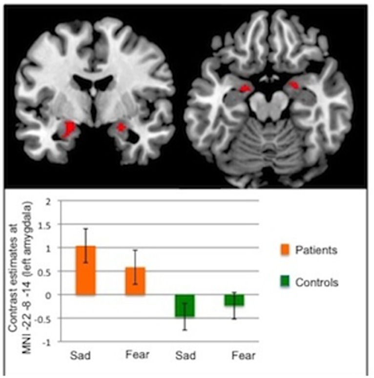
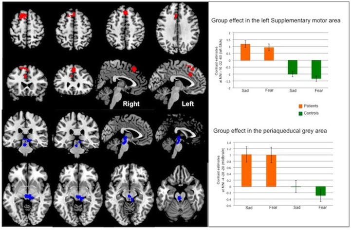
We found increased amygdala activation to negative emotions in CD compared to healthy controls in region of interest analyses, which persisted over time consistent with previous findings using emotional paradigms. Furthermore during whole brain analyses we found significantly increased activation in CD patients in areas involved in the 'freeze response' to fear (periaqueductal grey matter), and areas involved in self-awareness and motor control (cingulate gyrus and supplementary motor area).
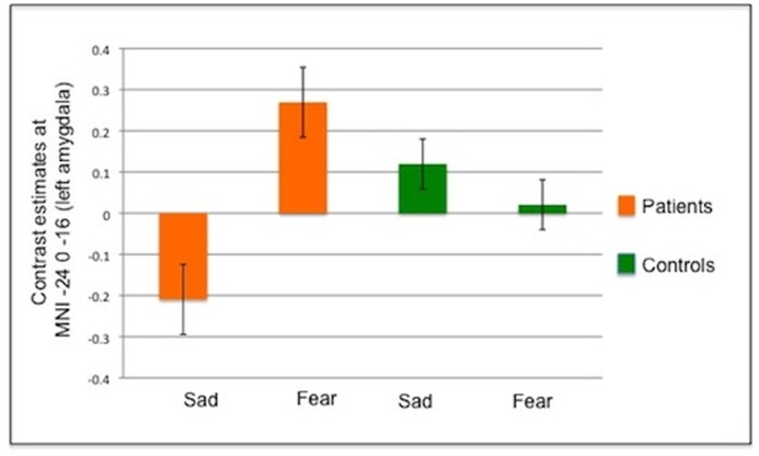
In contrast to healthy controls, CD patients exhibited increased response amplitude to fearful stimuli over time, suggesting abnormal emotional regulation (failure of habituation / sensitization). Patients with CD also activated midbrain and frontal structures that could reflect an abnormal behavioral-motor response to negative including threatening stimuli. This suggests a mechanism linking emotions to motor dysfunction in CD.
评估运动性转换障碍(CD)患者对负性情绪进行内隐加工的神经关联。
12名运动性CD患者和14名匹配的健康对照完成了一项事件相关功能磁共振成像(fMRI)任务,使用标准化的恐惧和悲伤表情面孔刺激与中性表情面孔进行对比。还对两组中刺激敏感性的时间变化进行了建模和测试。
在感兴趣区分析中,我们发现与健康对照相比,CD患者杏仁核对负性情绪的激活增加,且随时间持续存在,这与之前使用情绪范式的研究结果一致。此外,在全脑分析中,我们发现CD患者中参与对恐惧的“冻结反应”的区域(导水管周围灰质)以及参与自我意识和运动控制的区域(扣带回和辅助运动区)的激活显著增加。
与健康对照相比,CD患者对恐惧刺激的反应幅度随时间增加,提示情绪调节异常(习惯化/敏感化失败)。CD患者还激活了中脑和额叶结构,这可能反映了对包括威胁性刺激在内的负性刺激的异常行为运动反应。这提示了一种将情绪与CD中的运动功能障碍相联系的机制。