Abramson Richard G, Lambert Katrina F, Jones-Jackson Laurie B, Arlinghaus Lori R, Williams Jason, Abramson Vandana G, Chakravarthy A Bapsi, Yankeelov Thomas E
Department of Radiology and Radiological Sciences, Vanderbilt University, 1161 21st Avenue S, CCC-1121 MCN, Nashville, TN 37232-2675.
Department of Radiology and Radiological Sciences, Vanderbilt University, 1161 21st Avenue S, CCC-1121 MCN, Nashville, TN 37232-2675.
Acad Radiol. 2015 Jul;22(7):853-9. doi: 10.1016/j.acra.2015.02.012. Epub 2015 Apr 10.
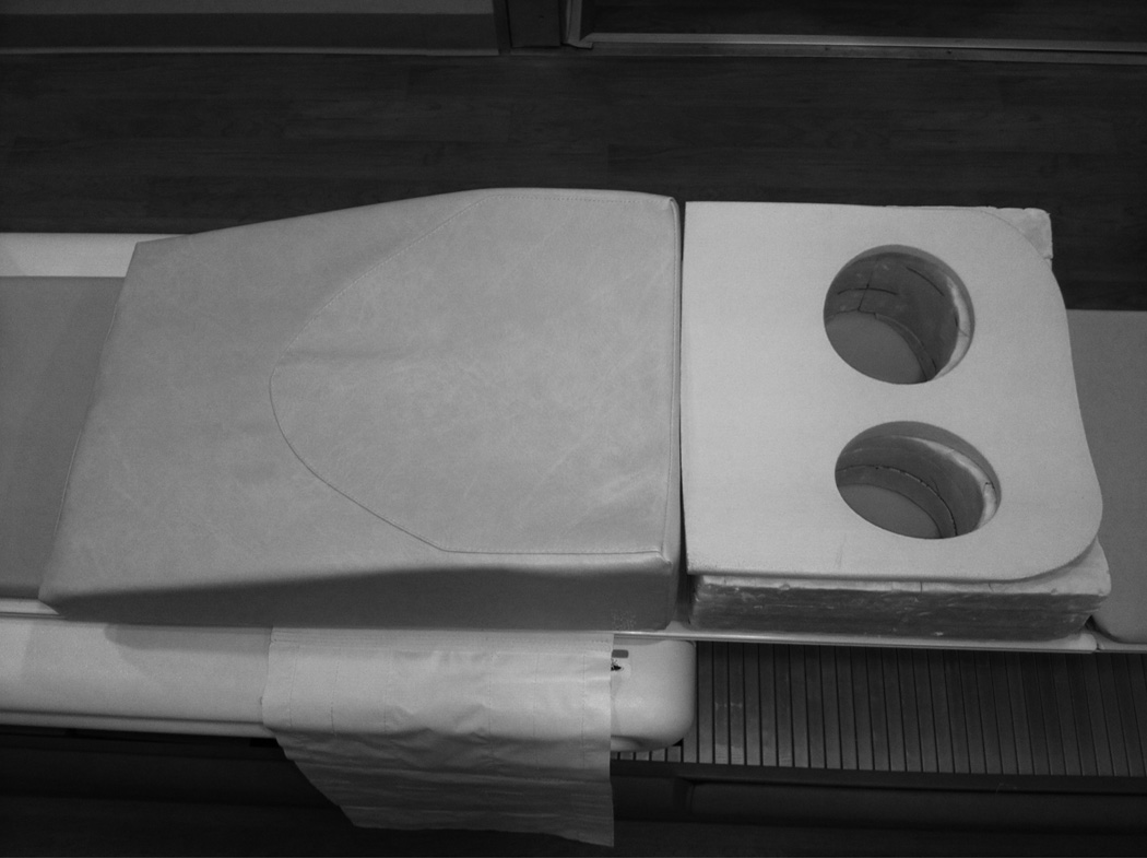
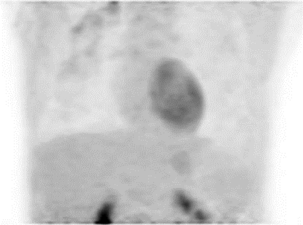
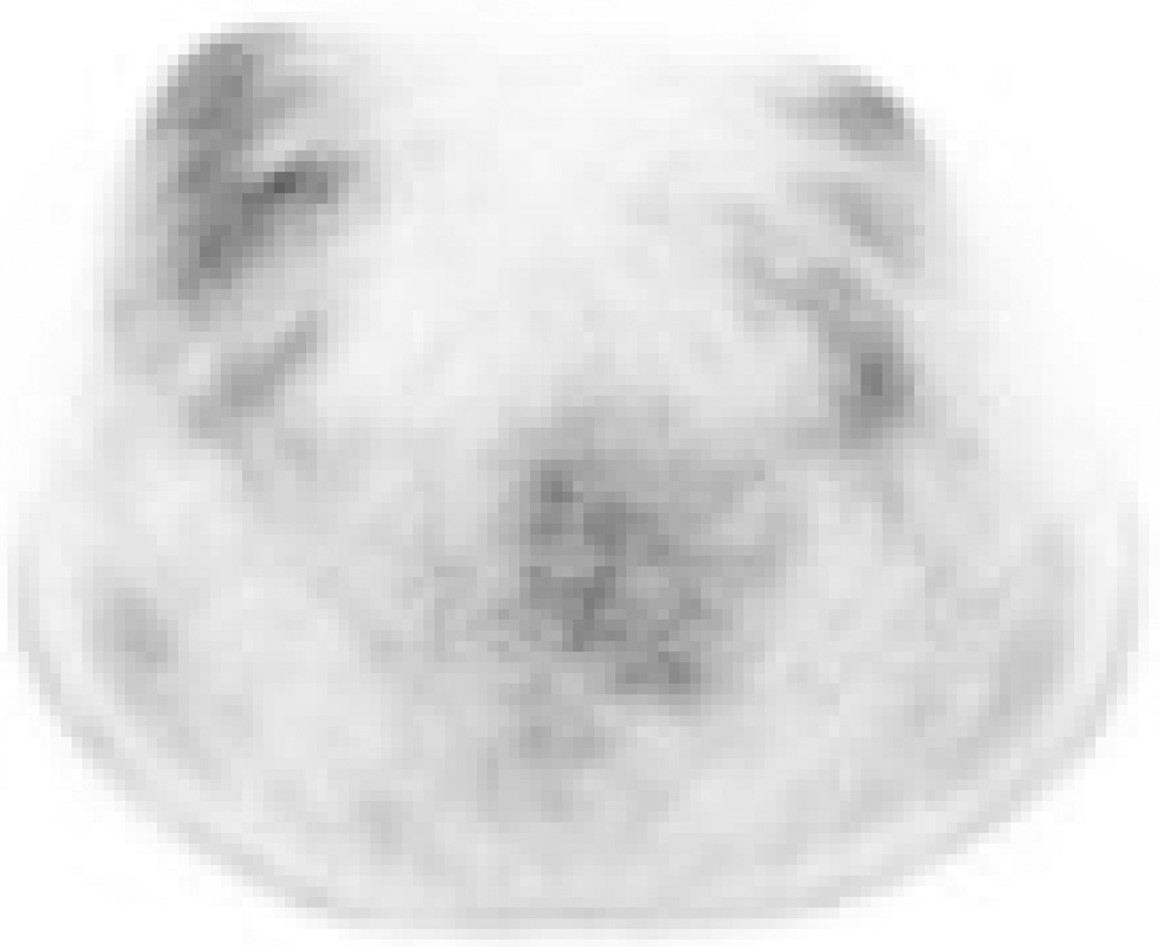
Prone (18)F fluorodeoxyglucose positron emission tomography/computed tomography (FDG-PET/CT) may have advantages for breast imaging because of improved separation of deep anatomic structures. There are limited data on whether prone and supine FDG-PET/CT provide similar information regarding breast and axillary disease in the setting of locally advanced breast cancer (LABC). The purpose of this study was to compare the information on locoregional disease distribution provided by prone versus supine FDG-PET in newly diagnosed LABC.
In an Institutional Review Board-approved prospective trial, 24 patients with newly diagnosed LABC underwent both supine and prone FDG-PET/CT at the same scanning session. Three readers performed an independent review of all scans and categorized the locoregional disease distribution as breast only (BO)-unifocal, BO-multifocal, BO-multicentric, or breast + axillary involvement. For breast + axillary disease, the readers also assessed the number of involved axillary lymph nodes. Interobserver discrepancies were resolved at a consensus reading session.
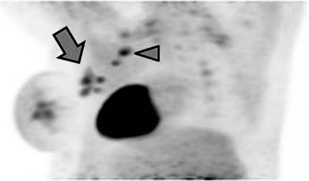
Two scanning sessions were excluded because the prone scan had omitted part of the axilla from the field of view. In the remaining 22 patients, the consensus categorization of anatomic disease distribution was concordant between prone and supine scanning in 21 patients (linear kappa 0.91, 95% confidence interval [0.79-1]). In the 16 patients with breast + axillary disease, equal numbers of involved lymph nodes were identified on prone and supine scanning in 12 patients, whereas in the remaining four patients, prone scanning resulted in a higher number of visualized lymph nodes.
Prone and supine FDG-PET/CT provided statistically identical information on locoregional disease distribution in LABC. However, prone scanning may perform better than supine for assessing the number of involved lymph nodes. Prone FDG-PET/CT may be useful in future clinical and research efforts, including hybrid PET-magnetic resonance imaging (MRI) applications.
俯卧位(18)F氟脱氧葡萄糖正电子发射断层扫描/计算机断层扫描(FDG-PET/CT)对乳腺成像可能具有优势,因为它能更好地分离深部解剖结构。关于在局部晚期乳腺癌(LABC)情况下,俯卧位和仰卧位FDG-PET/CT在乳腺和腋窝疾病方面是否提供相似信息的数据有限。本研究的目的是比较新诊断的LABC中俯卧位与仰卧位FDG-PET提供的关于局部区域疾病分布的信息。
在一项经机构审查委员会批准的前瞻性试验中,24例新诊断的LABC患者在同一扫描时段接受了仰卧位和俯卧位FDG-PET/CT检查。三位阅片者对所有扫描结果进行独立评估,并将局部区域疾病分布分类为仅乳腺(BO)-单灶性、BO-多灶性、BO-多中心性或乳腺+腋窝受累。对于乳腺+腋窝疾病,阅片者还评估了受累腋窝淋巴结的数量。观察者间的差异在共识读片会议上得到解决。
由于俯卧位扫描遗漏了腋窝的一部分视野,排除了两个扫描时段。在其余22例患者中,21例患者俯卧位和仰卧位扫描在解剖疾病分布的共识分类上是一致的(线性kappa值为0.91,95%置信区间[0.79 - 1])。在16例乳腺+腋窝疾病患者中,12例患者俯卧位和仰卧位扫描发现的受累淋巴结数量相等,而在其余4例患者中,俯卧位扫描发现的可见淋巴结数量更多。
俯卧位和仰卧位FDG-PET/CT在LABC的局部区域疾病分布方面提供了统计学上相同的信息。然而,在评估受累淋巴结数量方面,俯卧位扫描可能比仰卧位表现更好。俯卧位FDG-PET/CT可能在未来的临床和研究工作中有用,包括混合PET-磁共振成像(MRI)应用。