Halilovic Emina Alimanovic, Ljaljevic Sanida, Alimanovic Ilda, Mavija Milka, Oros Ana, Nisic Faruk
Eye Clinic, Clinical University Center Sarajevo, Bosnia and Herzegovina.
Eye Clinic, Clinical Center Banja Luka, Bosnia and Herzegovina.
Med Arch. 2015 Feb;69(1):34-7. doi: 10.5455/medarh.2015.69.34-37. Epub 2015 Feb 21.
Main the goal of the research is to analyze the occurrence of glaucoma in patients with diabetes mellitus type 1 (DM type 1) and diabetes mellitus type 2 (DM type 2).
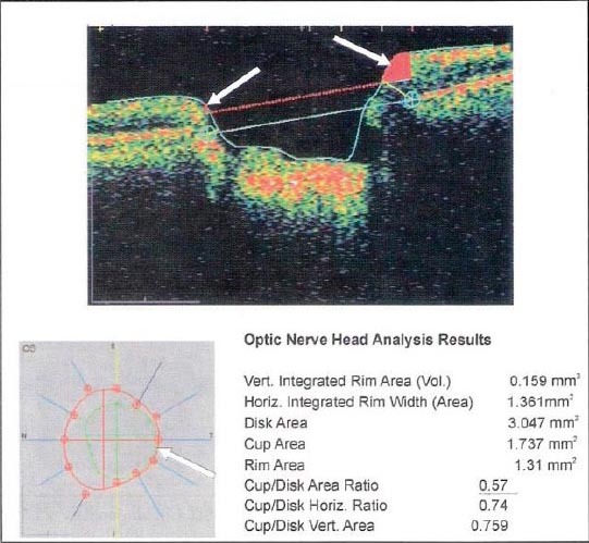
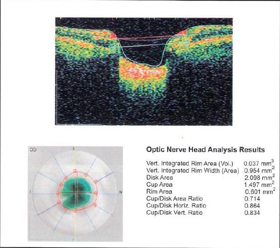
The study involved 140 patients, 34 with DM type 1 and 106 with DM type2. In relation to the type of glaucoma to the patients are divided into two groups: Primary and Secondary glaucoma. According to the stage of diabetic retinopathy (DR) patients were analyzed in three groups: non-proliferative, preproliferative and proliferative DR. Since ophthalmological parameters were analyzed: best corrected visual acuity (BCVA), intraocular pressure (IOP), visual field (VF) of computerized perimetry, excavatio optic nerve (E/D) by optic coherent tomography (OCT).
Applying the test of quotient chance found that subjects with DM type 1 have a 5.94 times greater chance of developing secondary glaucoma, but is of primary (P <0.0001). In patients with DM type 2, where the chance of getting the subjects of secondary glaucoma 4.43 times larger than that of the primary (P = 0.0002).
Patients with DM type have great chance of developing secondary glaucoma of the primary. Primary glaucoma more common in NPDR but secondary glaucoma more common in PDR.
本研究的主要目标是分析1型糖尿病(DM1)和2型糖尿病(DM2)患者青光眼的发生率。
该研究纳入了140例患者,其中34例为DM1患者,106例为DM2患者。根据青光眼类型,患者被分为两组:原发性青光眼和继发性青光眼。根据糖尿病视网膜病变(DR)的阶段,患者被分为三组进行分析:非增殖性、增殖前期和增殖性DR。分析的眼科参数包括:最佳矫正视力(BCVA)、眼压(IOP)、电脑视野计的视野(VF)、光学相干断层扫描(OCT)测量的视神经杯盘比(E/D)。
应用商数检验发现,DM1患者发生继发性青光眼的几率比原发性青光眼高5.94倍(P<0.0001)。在DM2患者中,发生继发性青光眼的几率比原发性青光眼高4.43倍(P=0.0002)。
DM患者发生继发性青光眼的几率高于原发性青光眼。原发性青光眼在非增殖性糖尿病视网膜病变中更常见,而继发性青光眼在增殖性糖尿病视网膜病变中更常见。