Li Xiaoguang, Zhu Yongshan, Kang Houyi, Zhang Yulong, Liang Huaping, Wang Sumei, Zhang Weiguo
Department of Radiology, Institute of Surgery Research, Daping Hospital, Third Military Medical University, Chongqing, 400042, China.
Department of Radiology, Tianchang people's hospital, Tianchang, Anhui, 239300, China.
Cancer Imaging. 2015 Mar 21;15(1):4. doi: 10.1186/s40644-015-0039-z.
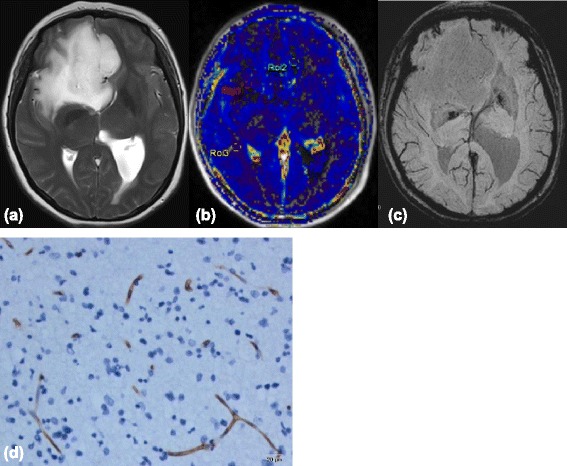
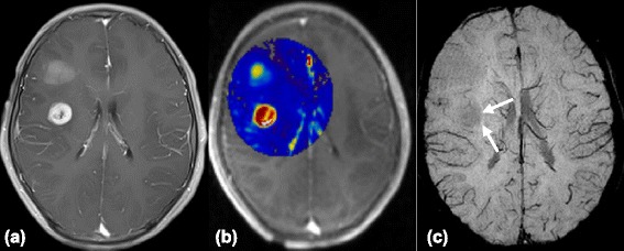
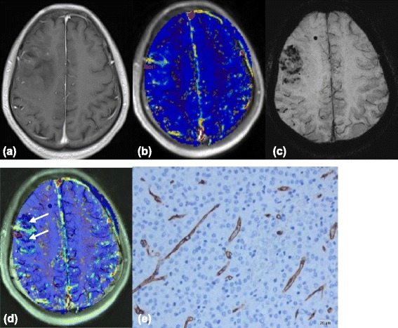
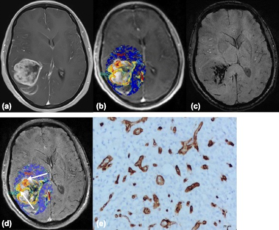
Dynamic contrast-enhanced MRI (DCE-MRI) estimates vascular permeability of brain tumors, and susceptibility-weighted imaging (SWI) may demonstrate tumor vascularity by intratumoral susceptibility signals (ITSS). This study assessed volume transfer constant (Ktrans) accuracy, the volume of extravascular extracellular space (EES) per unit volume of tissue (Ve) derived from DCE-MRI, and the degree of ITSS in glioma grading.
Thirty-two patients with different glioma grades were enrolled in this retrospective study. Patients underwent DCE-MRI and non-contrast enhanced SWI by three-tesla scanning. Ktrans values, Ve, and the degree of ITSS in glioma were compared. Receiver operating characteristic (ROC) curve analysis determined diagnostic performances of Ktrans and Ve in glioma grading, and Spearman's correlation analysis determined the associations between Ktrans, Ve, ITSS, and tumor grade.
Ktrans and Ve values were significantly different between low grade gliomas (LGGs) and both high grade gliomas (HGGs) and grade II, III and IV gliomas (P<0.01). The degree of ITSS of LGGs was lower than HGGs (P<0.01), and the ITSS of grade II gliomas was lower than grade III or IV gliomas. Ktrans and Ve were correlated with glioma grade (P<0.01), while ITSS was moderately correlated (P<0.01). Ktrans values were moderately correlated with ITSS in the same segments (P<0.01).
Ktrans and Ve values, and ITSS helped distinguish the differences between LGGs and HGGs and between grade II, III and IV gliomas. There was a moderate correlation between Ktrans and ITSS in the same tumor segments.
动态对比增强磁共振成像(DCE-MRI)可评估脑肿瘤的血管通透性,而磁敏感加权成像(SWI)可通过肿瘤内磁敏感信号(ITSS)显示肿瘤血管情况。本研究评估了容积转运常数(Ktrans)的准确性、从DCE-MRI得出的每单位组织体积的血管外细胞外间隙(EES)容积(Ve)以及胶质瘤分级中ITSS的程度。
本回顾性研究纳入了32例不同胶质瘤分级的患者。患者接受了3特斯拉扫描的DCE-MRI和非增强SWI检查。比较了胶质瘤的Ktrans值、Ve值和ITSS程度。采用受试者操作特征(ROC)曲线分析确定Ktrans和Ve在胶质瘤分级中的诊断性能,采用Spearman相关性分析确定Ktrans、Ve、ITSS与肿瘤分级之间的关联。
低级别胶质瘤(LGG)与高级别胶质瘤(HGG)以及II、III和IV级胶质瘤之间的Ktrans值和Ve值存在显著差异(P<0.01)。LGG的ITSS程度低于HGG(P<0.01),II级胶质瘤的ITSS低于III级或IV级胶质瘤。Ktrans和Ve与胶质瘤分级相关(P<0.01),而ITSS呈中度相关(P<0.01)。同一节段的Ktrans值与ITSS呈中度相关(P<0.01)。
Ktrans值、Ve值和ITSS有助于区分LGG与HGG以及II、III和IV级胶质瘤之间的差异。同一肿瘤节段的Ktrans与ITSS之间存在中度相关性。