Kozor Rebecca, Callaghan Fraser, Tchan Michel, Hamilton-Craig Christian, Figtree Gemma A, Grieve Stuart M
North Shore Heart Research Group, Kolling Institute of Medical Research, University of Sydney, Sydney, Australia.
Department of Cardiology, Royal North Shore Hospital, Sydney, Australia.
J Cardiovasc Magn Reson. 2015 Feb 21;17(1):22. doi: 10.1186/s12968-015-0114-4.
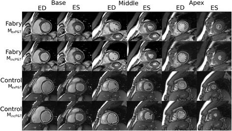
Sphingolipid deposition in Fabry disease causes left ventricular (LV) hypertrophy, of which the accurate assessment is essential. Cardiovascular magnetic resonance (CMR) has been proposed as the gold standard. However, there is debate in the literature as to whether papillary muscles and trabeculations (P&T) should be included in LV mass (LVM).
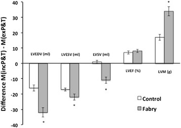
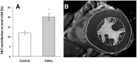
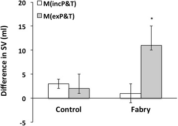
METHODS/RESULTS: We examined the accuracy of 2 CMR methods of assessing LVM and LV volumes, including (M inc P&T) or excluding (M ex P&T) P&T, in a cohort of Fabry disease subjects (n = 20) compared to a matched control group (n = 20). Significant differences between the two measurement methods were observed for LV end-diastolic volume, LV end-systolic volume, LVM, and LV ejection fraction (LVEF) in both groups. These differences were significantly greater in the Fabry group compared to controls, except for LVEF. P&T contributed to a greater percentage of LVM in Fabry subjects than controls (20 ± 1% vs 13 ± 2%, p = 0.01). In the control group, both volume-derived methods (M inc P&T or MexP&T) provided accurate SV measurements compared with the internal reference of velocity-encoded aortic flow. In the Fabry group, inclusion of P&T (M inc P&T) resulted in good concordance with phase contrast flow imaging (difference between flow and volume techniques: 1 ± 3 ml, p = 0.7).
The volumetric contribution of P&T in Fabry disease is markedly increased relative to healthy controls. Failure to account for this results in significant underestimation of LVM and results in misclassification of a proportion of subjects.
法布里病中鞘脂沉积会导致左心室(LV)肥厚,准确评估至关重要。心血管磁共振成像(CMR)已被提议作为金标准。然而,文献中对于乳头肌和小梁(P&T)是否应包含在左心室质量(LVM)中存在争议。
方法/结果:我们在一组法布里病受试者(n = 20)中,与匹配的对照组(n = 20)相比,研究了两种评估LVM和左心室容积的CMR方法的准确性,这两种方法分别是包含(M inc P&T)或排除(M ex P&T)P&T。两组在左心室舒张末期容积、左心室收缩末期容积、LVM和左心室射血分数(LVEF)方面,两种测量方法均观察到显著差异。除LVEF外,法布里病组的这些差异比对照组显著更大。与对照组相比,法布里病受试者中P&T对LVM的贡献百分比更高(20±1%对13±2%,p = 0.01)。在对照组中,与速度编码主动脉血流的内部参考相比,两种基于容积的方法(M inc P&T或MexP&T)均提供了准确的每搏输出量(SV)测量值。在法布里病组中,包含P&T(M inc P&T)与相位对比血流成像具有良好的一致性(血流与容积技术之间的差异:1±3 ml,p = 0.7)。
相对于健康对照组,法布里病中P&T的容积贡献显著增加。未考虑这一点会导致LVM被显著低估,并导致一部分受试者被错误分类。