• 文献检索
  • 文档翻译
  • 深度研究
  • 学术资讯
  • Suppr Zotero 插件Zotero 插件
  • 邀请有礼
  • 套餐&价格
  • 历史记录
应用&插件
Suppr Zotero 插件Zotero 插件浏览器插件Mac 客户端Windows 客户端微信小程序
定价
高级版会员购买积分包购买API积分包
服务
文献检索文档翻译深度研究API 文档MCP 服务
关于我们
关于 Suppr公司介绍联系我们用户协议隐私条款
关注我们

Suppr 超能文献

核心技术专利:CN118964589B侵权必究
粤ICP备2023148730 号-1Suppr @ 2026

文献检索

告别复杂PubMed语法,用中文像聊天一样搜索,搜遍4000万医学文献。AI智能推荐,让科研检索更轻松。

立即免费搜索

文件翻译

保留排版,准确专业,支持PDF/Word/PPT等文件格式,支持 12+语言互译。

免费翻译文档

深度研究

AI帮你快速写综述,25分钟生成高质量综述,智能提取关键信息,辅助科研写作。

立即免费体验

心血管磁共振长轴和短轴左心室容积参数定量比较及其与离体验证。

Comparison of long and short axis quantification of left ventricular volume parameters by cardiovascular magnetic resonance, with ex-vivo validation.

机构信息

Stephenson Cardiovascular MR Centre at Libin Cardiovascular Institute of Alberta, Departments of Cardiac Sciences and Radiology, University of Calgary, AB, Canada.

出版信息

J Cardiovasc Magn Reson. 2011 Aug 11;13(1):40. doi: 10.1186/1532-429X-13-40.

DOI:10.1186/1532-429X-13-40
PMID:21834992
原文链接:https://pmc.ncbi.nlm.nih.gov/articles/PMC3169477/
Abstract

BACKGROUND

The purpose of the study was to compare the accuracy and evaluation time of quantifying left ventricular (LV), left atrial (LA) volume and LV mass using short axis (SAX) and long axis (LAX) methods when using cardiovascular magnetic resonance (CMR).

MATERIALS AND METHODS

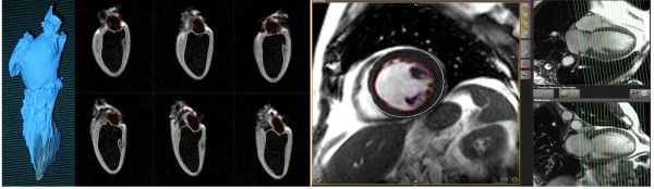
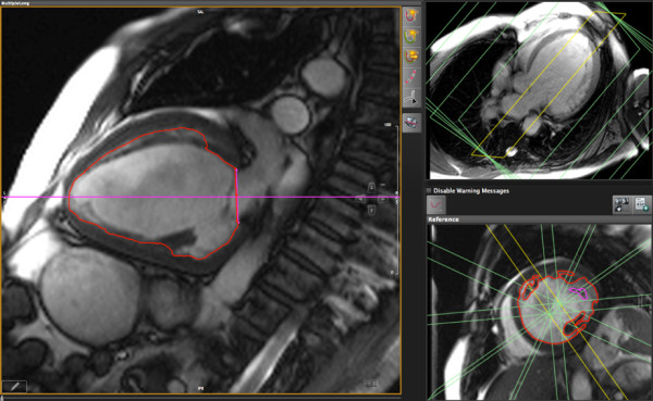
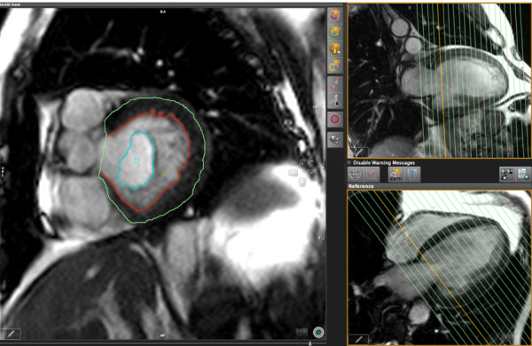
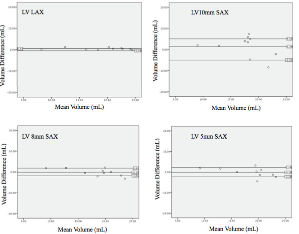
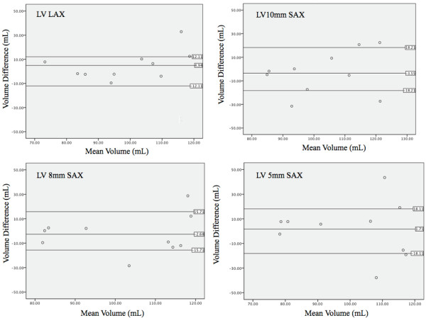
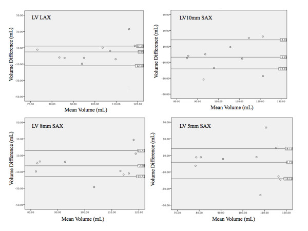
We studied 12 explanted canine hearts and 46 patients referred for CMR (29 male, age 47 ± 18 years) in a clinical 1.5 T CMR system, using standard cine sequences. In standard short axis stacks of various slice thickness values in dogs and 8 mm slice thickness (gap 2 mm) in patients, we measured LV volumes using reference slices in a perpendicular, long axis orientation using certified software. Volumes and mass were also measured in six radial long axis (LAX) views.LV parameters were also assessed for intra- and inter-observer variability. In 24 patients, we also analyzed reproducibility and evaluation time of two very experienced (> 10 years of CMR reading) readers for SAX and LAX.

RESULTS

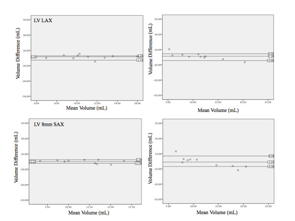
In the explanted dog hearts, there was excellent agreement between ex vivo data and LV mass and volume data as measured by all methods for both, LAX (r² = 0.98) and SAX (r² = 0.88 to 0.98). LA volumes, however, were underestimated by 13% using the LAX views. In patients, there was a good correlation between all three assessed methods (r² ≥ 0.95 for all). In experienced clinical readers, left-ventricular volumes and ejection fraction as measured in LAX views showed a better inter-observer reproducibility and a 27% shorter evaluation time.

CONCLUSION

When compared to an ex vivo standard, both, short axis and long axis techniques are highly accurate for the quantification of left ventricular volumes and mass. In clinical settings, however, the long axis approach may be more reproducible and more time-efficient. Therefore, the rotational long axis approach is a viable alternative for the clinical assessment of cardiac volumes, function and mass.

摘要

背景

本研究旨在比较心血管磁共振(CMR)中使用短轴(SAX)和长轴(LAX)方法定量左心室(LV)、左心房(LA)容积和 LV 质量的准确性和评估时间。

材料和方法

我们在临床 1.5T CMR 系统中研究了 12 个离体犬心和 46 例因 CMR 就诊的患者(29 名男性,年龄 47±18 岁),使用标准电影序列。在犬的不同切片厚度值的标准短轴堆栈和患者的 8mm 切片厚度(间隙 2mm)中,我们使用垂直长轴方向的参考切片,使用认证软件测量 LV 容积。还在六个径向长轴(LAX)视图中测量了容积和质量。还评估了 LV 参数的观察者内和观察者间变异性。在 24 例患者中,我们还分析了两位经验丰富(CMR 阅读超过 10 年)的读者对 SAX 和 LAX 的重复性和评估时间。

结果

在离体犬心,LAX(r²=0.98)和 SAX(r²=0.88 至 0.98)所有方法测量的离体数据与 LV 质量和容积数据之间存在极好的一致性。然而,LAX 视图低估了 LA 容积 13%。在患者中,所有三种评估方法之间存在良好的相关性(r²≥0.95)。在经验丰富的临床读者中,LAX 视图中测量的左心室容积和射血分数具有更好的观察者间可重复性,评估时间缩短了 27%。

结论

与离体标准相比,短轴和长轴技术均高度准确地定量左心室容积和质量。然而,在临床环境中,长轴方法可能更具可重复性和更高效。因此,旋转长轴方法是评估心脏容积、功能和质量的一种可行替代方法。

https://cdn.ncbi.nlm.nih.gov/pmc/blobs/7c0c/3169477/36a4f21f39da/1532-429X-13-40-7.jpg
https://cdn.ncbi.nlm.nih.gov/pmc/blobs/7c0c/3169477/0080e7d5cd22/1532-429X-13-40-1.jpg
https://cdn.ncbi.nlm.nih.gov/pmc/blobs/7c0c/3169477/b64ee7b6f19a/1532-429X-13-40-2.jpg
https://cdn.ncbi.nlm.nih.gov/pmc/blobs/7c0c/3169477/d09992e767ed/1532-429X-13-40-3.jpg
https://cdn.ncbi.nlm.nih.gov/pmc/blobs/7c0c/3169477/ea406f7ad3ab/1532-429X-13-40-4.jpg
https://cdn.ncbi.nlm.nih.gov/pmc/blobs/7c0c/3169477/0f67136c2531/1532-429X-13-40-5.jpg
https://cdn.ncbi.nlm.nih.gov/pmc/blobs/7c0c/3169477/6784b83c44cd/1532-429X-13-40-6.jpg
https://cdn.ncbi.nlm.nih.gov/pmc/blobs/7c0c/3169477/36a4f21f39da/1532-429X-13-40-7.jpg

相似文献

1
Comparison of long and short axis quantification of left ventricular volume parameters by cardiovascular magnetic resonance, with ex-vivo validation.心血管磁共振长轴和短轴左心室容积参数定量比较及其与离体验证。
J Cardiovasc Magn Reson. 2011 Aug 11;13(1):40. doi: 10.1186/1532-429X-13-40.
2
Compressed sensing single-breath-hold CMR for fast quantification of LV function, volumes, and mass.压缩感知单次屏气心脏磁共振快速定量左心室功能、容积和质量。
JACC Cardiovasc Imaging. 2014 Sep;7(9):882-92. doi: 10.1016/j.jcmg.2014.04.016. Epub 2014 Aug 13.
3
Transthoracic 3D Echocardiographic Left Heart Chamber Quantification Using an Automated Adaptive Analytics Algorithm.经胸三维超声心动图左心腔定量分析的自动自适应分析算法。
JACC Cardiovasc Imaging. 2016 Jul;9(7):769-782. doi: 10.1016/j.jcmg.2015.12.020. Epub 2016 Jun 15.
4
Biplane versus short-axis measures of the left atrium and ventricle in patients with systolic dysfunction assessed by magnetic resonance.磁共振评估收缩功能不全患者时左心房和左心室的双平面与短轴测量
Clin Imaging. 2016 Sep-Oct;40(5):907-12. doi: 10.1016/j.clinimag.2016.04.015. Epub 2016 Apr 30.
5
Compressed sensing real-time cine cardiovascular magnetic resonance: accurate assessment of left ventricular function in a single-breath-hold.压缩感知实时电影心血管磁共振成像:单次屏气下对左心室功能的准确评估
J Cardiovasc Magn Reson. 2016 Aug 24;18(1):50. doi: 10.1186/s12968-016-0271-0.
6
Diagnosis of Left Ventricular Diastolic Dysfunction Using Cardiac Magnetic Resonance Imaging: Comparison of Volume-Time Curves Derived from Long- and Short-Axis Cine Steady-State Free Precession Datasets.应用心脏磁共振成像诊断左心室舒张功能障碍:长轴和短轴电影稳态自由进动数据集容积-时间曲线的比较。
Rofo. 2020 Aug;192(8):764-775. doi: 10.1055/a-1108-1892. Epub 2020 Feb 27.
7
The intra-observer reproducibility of cardiovascular magnetic resonance myocardial feature tracking strain assessment is independent of field strength.心血管磁共振心肌特征追踪应变评估的观察者内可重复性与磁场强度无关。
Eur J Radiol. 2013 Feb;82(2):296-301. doi: 10.1016/j.ejrad.2012.11.012. Epub 2012 Dec 12.
8
Single breath-hold 3D measurement of left atrial volume using compressed sensing cardiovascular magnetic resonance and a non-model-based reconstruction approach.使用压缩感知心血管磁共振成像和基于非模型的重建方法对左心房容积进行单次屏气三维测量。
J Cardiovasc Magn Reson. 2015 Jun 11;17(1):47. doi: 10.1186/s12968-015-0147-8.
9
Inter-study reproducibility of cardiovascular magnetic resonance myocardial feature tracking.心血管磁共振心肌特征追踪研究间可重复性。
J Cardiovasc Magn Reson. 2012 Jun 21;14(1):43. doi: 10.1186/1532-429X-14-43.
10
Accelerating global left-ventricular function assessment in mice using reduced slice acquisition and three-dimensional guide-point modelling.使用减少切片采集和三维引导点建模技术加速小鼠的全球左心室功能评估。
J Cardiovasc Magn Reson. 2011 Sep 14;13(1):49. doi: 10.1186/1532-429X-13-49.

引用本文的文献

1
Development and validation of AI-derived segmentation of four-chamber cine cardiac magnetic resonance.四腔心电影心脏磁共振人工智能分割的研发与验证。
Eur Radiol Exp. 2024 Jul 12;8(1):77. doi: 10.1186/s41747-024-00477-7.
2
Comparative analysis of left ventricle function and deformation imaging in short and long axis plane in cardiac magnetic resonance imaging.心脏磁共振成像中短轴和长轴平面左心室功能与形变成像的对比分析
Front Cardiovasc Med. 2024 May 2;11:1388171. doi: 10.3389/fcvm.2024.1388171. eCollection 2024.
3
The additive prognostic value of end-systolic pressure-volume relation by stress CMR in patients with known or suspected coronary artery disease.

本文引用的文献

1
ACCF/ACR/AHA/NASCI/SCMR 2010 expert consensus document on cardiovascular magnetic resonance: a report of the American College of Cardiology Foundation Task Force on Expert Consensus Documents.ACCF/ACR/AHA/NASCI/SCMR 2010年心血管磁共振专家共识文件:美国心脏病学会基金会专家共识文件特别工作组报告
Circulation. 2010 Jun 8;121(22):2462-508. doi: 10.1161/CIR.0b013e3181d44a8f. Epub 2010 May 17.
2
Utility of cardiovascular magnetic resonance to predict left ventricular recovery after primary percutaneous coronary intervention for patients presenting with acute ST-segment elevation myocardial infarction.心血管磁共振成像在预测急性ST段抬高型心肌梗死患者接受直接经皮冠状动脉介入治疗后左心室恢复情况中的应用
Am J Cardiol. 2007 Jul 15;100(2):211-6. doi: 10.1016/j.amjcard.2007.02.079. Epub 2007 Jun 4.
3
应力心脏磁共振成像测定的收缩末期压力-容积关系在已知或疑似冠心病患者中的附加预后价值。
Int J Cardiovasc Imaging. 2024 Jun;40(6):1341-1351. doi: 10.1007/s10554-024-03104-z. Epub 2024 Apr 27.
4
An artificial intelligence tool for automated analysis of large-scale unstructured clinical cine cardiac magnetic resonance databases.一种用于自动分析大规模非结构化临床心脏磁共振电影数据库的人工智能工具。
Eur Heart J Digit Health. 2023 Jul 13;4(5):370-383. doi: 10.1093/ehjdh/ztad044. eCollection 2023 Oct.
5
Cardiovascular magnetic resonance-derived left atrioventricular coupling index and major adverse cardiac events in patients following acute myocardial infarction.心血管磁共振衍生的左心房-左心室偶联指数与急性心肌梗死后患者的主要不良心脏事件。
J Cardiovasc Magn Reson. 2023 Apr 13;25(1):24. doi: 10.1186/s12968-023-00929-w.
6
Influence of MRI-based boundary conditions on type B aortic dissection simulations in false lumen with or without abdominal aorta involvement.基于磁共振成像(MRI)的边界条件对伴有或不伴有腹主动脉受累的B型主动脉夹层假腔模拟的影响。
Front Physiol. 2022 Sep 7;13:977275. doi: 10.3389/fphys.2022.977275. eCollection 2022.
7
The role of cardiac magnetic resonance imaging in the assessment of heart failure with preserved ejection fraction.心脏磁共振成像在射血分数保留的心力衰竭评估中的作用。
Front Cardiovasc Med. 2022 Jul 18;9:922398. doi: 10.3389/fcvm.2022.922398. eCollection 2022.
8
Cerebrovascular Variants and the Role of the Selfish Brain in Young-Onset Hypertension.脑血管变异与青年期高血压中“自私大脑”的作用
Hypertension. 2022 Jun;79(6):1265-1274. doi: 10.1161/HYPERTENSIONAHA.121.18612. Epub 2022 Mar 16.
9
Artificial intelligence can detect left ventricular dilatation on contrast-enhanced thoracic computer tomography relative to cardiac magnetic resonance imaging.人工智能可以在对比增强胸部计算机断层扫描上检测到左心室扩张,相对于心脏磁共振成像。
Br J Radiol. 2022 Sep 1;95(1138):20210852. doi: 10.1259/bjr.20210852. Epub 2022 Mar 18.
10
Normal sex and age-specific parameters in a multi-ethnic population: a cardiovascular magnetic resonance study of the Canadian Alliance for Healthy Hearts and Minds cohort.多民族人群中的正常性别和年龄特异性参数:加拿大心脏和思维健康联盟队列的心血管磁共振研究。
J Cardiovasc Magn Reson. 2022 Jan 3;24(1):2. doi: 10.1186/s12968-021-00819-z.
ACCF/ACR/SCCT/SCMR/ASNC/NASCI/SCAI/SIR 2006 appropriateness criteria for cardiac computed tomography and cardiac magnetic resonance imaging: a report of the American College of Cardiology Foundation Quality Strategic Directions Committee Appropriateness Criteria Working Group, American College of Radiology, Society of Cardiovascular Computed Tomography, Society for Cardiovascular Magnetic Resonance, American Society of Nuclear Cardiology, North American Society for Cardiac Imaging, Society for Cardiovascular Angiography and Interventions, and Society of Interventional Radiology.美国心脏病学会基金会/美国放射学会/心血管计算机断层扫描学会/心血管磁共振学会/美国核医学会/北美心脏成像学会/心血管造影和介入学会/介入放射学会2006年心脏计算机断层扫描和心脏磁共振成像适宜性标准:美国心脏病学会基金会质量战略方向委员会适宜性标准工作组、美国放射学会、心血管计算机断层扫描学会、心血管磁共振学会、美国核医学会、北美心脏成像学会、心血管造影和介入学会以及介入放射学会的报告。
J Am Coll Cardiol. 2006 Oct 3;48(7):1475-97. doi: 10.1016/j.jacc.2006.07.003.
4
Normal human left and right ventricular and left atrial dimensions using steady state free precession magnetic resonance imaging.使用稳态自由进动磁共振成像测量正常人体左、右心室及左心房尺寸。
J Cardiovasc Magn Reson. 2005;7(5):775-82. doi: 10.1080/10976640500295516.
5
Effect of endocardial trabeculae on left ventricular measurements and measurement reproducibility at cardiovascular MR imaging.心内膜小梁对心血管磁共振成像中左心室测量及测量可重复性的影响。
Radiology. 2005 Jul;236(1):57-64. doi: 10.1148/radiol.2353040601. Epub 2005 Jun 13.
6
Left ventricular mass: manual and automatic segmentation of true FISP and FLASH cine MR images in dogs and pigs.左心室质量:犬类和猪类中真实稳态自由进动序列(true FISP)和快速小角度激发序列(FLASH)心脏电影磁共振成像的手动和自动分割
Radiology. 2004 Feb;230(2):389-95. doi: 10.1148/radiol.2302020761. Epub 2003 Dec 29.
7
Cine MRI using steady state free precession in the radial long axis orientation is a fast accurate method for obtaining volumetric data of the left ventricle.采用稳态自由进动序列在径向长轴方向进行的心脏磁共振成像(Cine MRI)是一种快速准确获取左心室容积数据的方法。
J Magn Reson Imaging. 2001 Dec;14(6):685-92. doi: 10.1002/jmri.10019.
8
The diagnostic impact of 2D- versus 3D- left ventricular volumetry by MRI in patients with suspected heart failure.心脏磁共振成像二维与三维左心室容积测定对疑似心力衰竭患者的诊断影响
MAGMA. 2000 Nov;11(1-2):16-9. doi: 10.1007/BF02678483.
9
Global cardiac function using fast breath-hold MRI: validation of new acquisition and analysis techniques.
Magn Reson Med. 1997 May;37(5):683-92. doi: 10.1002/mrm.1910370510.
10
Quantification of the left ventricular volumes and function with cine MR imaging: comparison of geometric models with three-dimensional data.采用电影磁共振成像定量分析左心室容积和功能:几何模型与三维数据的比较
Radiology. 1993 Aug;188(2):371-6. doi: 10.1148/radiology.188.2.8327680.