Škrášková Karolina, Khmelinskii Artem, Abdelmoula Walid M, De Munter Stephanie, Baes Myriam, McDonnell Liam, Dijkstra Jouke, Heeren Ron M A
FOM-Institute AMOLF, Amsterdam, The Netherlands.
J Am Soc Mass Spectrom. 2015 Jun;26(6):948-57. doi: 10.1007/s13361-015-1146-6. Epub 2015 Apr 28.
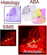
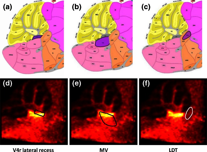
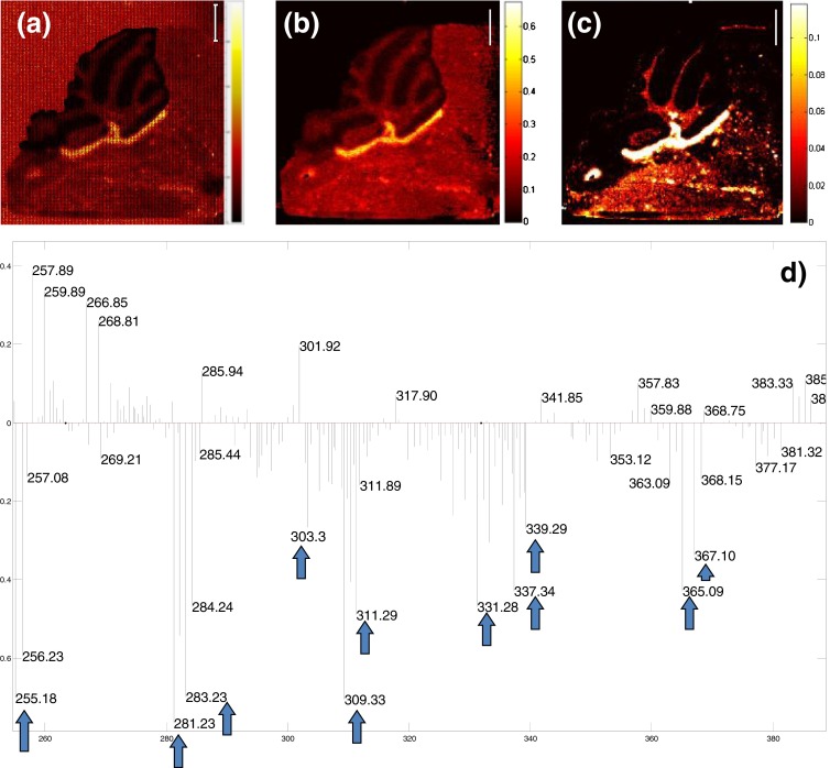
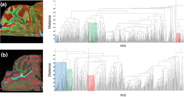
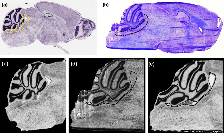
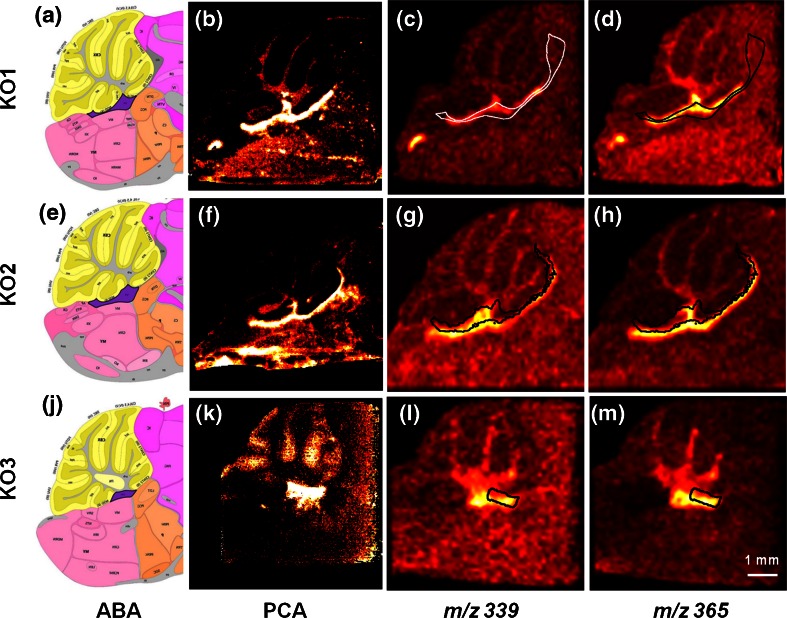
Mass spectrometry imaging (MSI) is a powerful tool for the molecular characterization of specific tissue regions. Histochemical staining provides anatomic information complementary to MSI data. The combination of both modalities has been proven to be beneficial. However, direct comparison of histology based and mass spectrometry-based molecular images can become problematic because of potential tissue damages or changes caused by different sample preparation. Curated atlases such as the Allen Brain Atlas (ABA) offer a collection of highly detailed and standardized anatomic information. Direct comparison of MSI brain data to the ABA allows for conclusions to be drawn on precise anatomic localization of the molecular signal. Here we applied secondary ion mass spectrometry imaging at high spatial resolution to study brains of knock-out mouse models with impaired peroxisomal β-oxidation. Murine models were lacking D-multifunctional protein (MFP2), which is involved in degradation of very long chain fatty acids. SIMS imaging revealed deposits of fatty acids within distinct brain regions. Manual comparison of the MSI data with the histologic stains did not allow for an unequivocal anatomic identification of the fatty acids rich regions. We further employed an automated pipeline for co-registration of the SIMS data to the ABA. The registration enabled precise anatomic annotation of the brain structures with the revealed lipid deposits. The precise anatomic localization allowed for a deeper insight into the pathology of Mfp2 deficient mouse models.
质谱成像(MSI)是对特定组织区域进行分子表征的强大工具。组织化学染色提供与MSI数据互补的解剖学信息。两种方法的结合已被证明是有益的。然而,由于不同样品制备可能导致的潜在组织损伤或变化,基于组织学和基于质谱的分子图像的直接比较可能会出现问题。诸如艾伦脑图谱(ABA)等精心策划的图谱提供了高度详细和标准化的解剖学信息集合。将MSI脑数据与ABA进行直接比较,可以得出分子信号精确解剖定位的结论。在这里,我们应用高空间分辨率的二次离子质谱成像来研究过氧化物酶体β-氧化受损的基因敲除小鼠模型的大脑。小鼠模型缺乏参与极长链脂肪酸降解的D-多功能蛋白(MFP2)。二次离子质谱成像揭示了不同脑区脂肪酸的沉积。将MSI数据与组织学染色进行人工比较,无法明确鉴定富含脂肪酸的区域。我们进一步采用了一种自动流程,将二次离子质谱数据与ABA进行配准。配准实现了对显示脂质沉积的脑结构的精确解剖标注。精确的解剖定位有助于更深入地了解Mfp2缺陷小鼠模型的病理学。