Zhu Yanbei, Yang Rongze, McLenithan John, Yu Daozhan, Wang Hong, Wang Yaping, Singh Devinder, Olson John, Sztalryd Carole, Zhu Dalong, Gong Da-Wei
Medical School of Nanjing University, Nanjing, China; Division of Endocrinology, Diabetes and Nutrition, Department of Medicine, University of Maryland School of Medicine, Baltimore, USA.
Obesity (Silver Spring). 2015 May;23(5):1014-21. doi: 10.1002/oby.21062.
To determine whether super-activation of PPARγ can reprogram human myoblasts into brown-like adipocytes and to establish a new cell model for browning research.
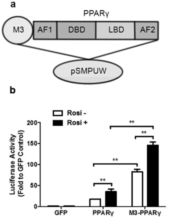
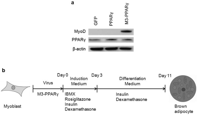
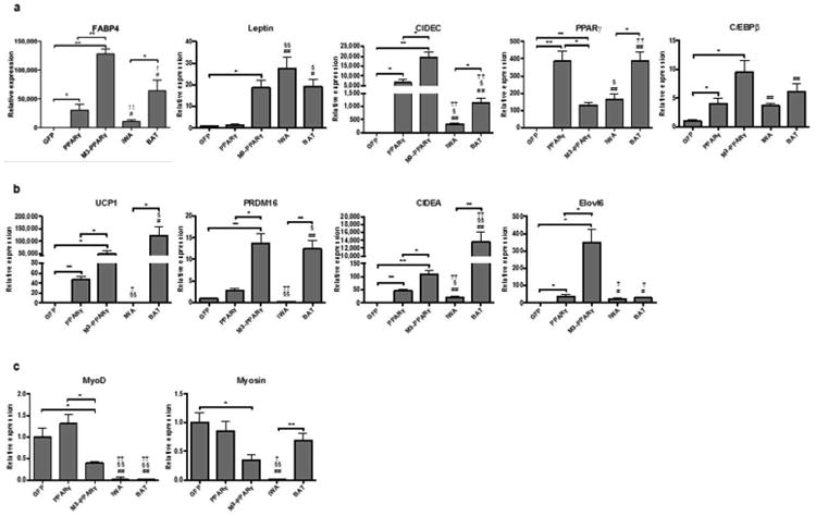
To enhance the PPARγ signaling, M3, the transactivation domain of MyoD, was fused to PPARγ. PPARγ and M3-PPARγ-lentiviral vectors were used to convert human myoblasts into adipocytes. Brown adipocyte markers of the reprogrammed adipocytes were assessed by qPCR and protein analyses. White adipocytes differentiated from subcutaneous stromal vascular cells and perithyroid brown fat tissues were used as references.
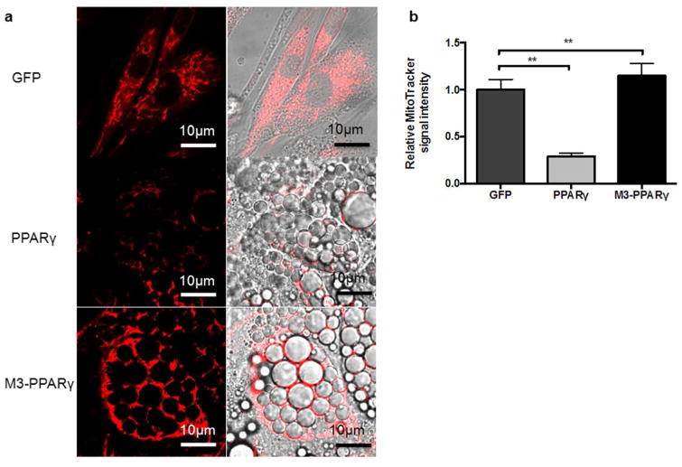
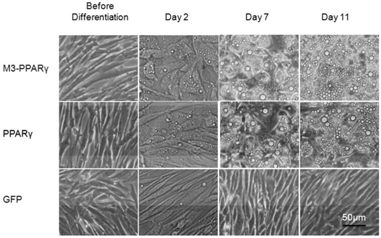
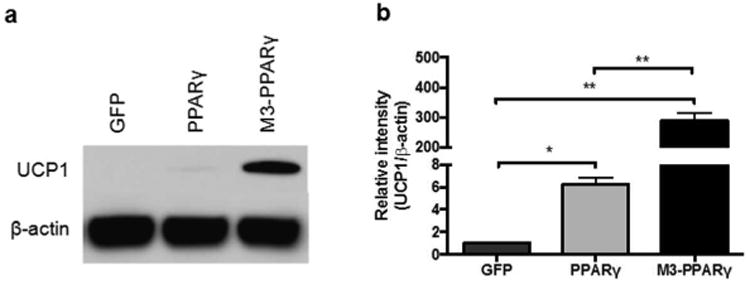
In transient transfections, M3-PPARγ had a stronger constitutive activity than PPARγ by reporter assay. Although the transduction of either PPARγ or M3-PPARγ induced adipogenesis in myoblasts, M3-PPARγ drastically induced the brown adipocyte markers of UCP1, CIDEA, and PRDM16 by 1,050, 2.4, and 5.0 fold, respectively and increased mitochondria contents by 4 fold, compared to PPARγ.
Super-activation of PPARγ can effectively convert human myoblasts into brown-like adipocytes and a new approach to derive brown-like adipocytes.
确定过氧化物酶体增殖物激活受体γ(PPARγ)的超激活是否能将人成肌细胞重编程为褐色脂肪样细胞,并建立一种用于褐色化研究的新细胞模型。
为增强PPARγ信号传导,将肌分化蛋白(MyoD)的反式激活结构域M3与PPARγ融合。使用PPARγ和M3-PPARγ慢病毒载体将人成肌细胞转化为脂肪细胞。通过定量聚合酶链反应(qPCR)和蛋白质分析评估重编程脂肪细胞的褐色脂肪细胞标志物。将从皮下基质血管细胞和甲状腺周围褐色脂肪组织分化而来的白色脂肪细胞用作对照。
在瞬时转染中,通过报告基因检测,M3-PPARγ具有比PPARγ更强的组成活性。尽管PPARγ或M3-PPARγ的转导均诱导成肌细胞发生脂肪生成,但与PPARγ相比,M3-PPARγ分别将解偶联蛋白1(UCP1)、细胞死亡诱导DFFA样效应因子A(CIDEA)和含锌指结构域的PR结构域蛋白16(PRDM16)的褐色脂肪细胞标志物大幅诱导了1050倍、2.4倍和5.0倍,并使线粒体含量增加了4倍。
PPARγ的超激活可有效将人成肌细胞转化为褐色脂肪样细胞,这是一种获得褐色脂肪样细胞的新方法。