Chavan Vrushali, Willis Jeffery, Walker Sidney K, Clark Helen R, Liu Xinran, Fox Michael A, Srivastava Sarika, Mukherjee Konark
Virginia Tech Carilion Research Institute, Roanoke, VA, 24016, United States of America.
Yale University, School of Medicine, Department of Cell Biology, New Haven, CT, 06510, United States of America.
PLoS One. 2015 Apr 30;10(4):e0125185. doi: 10.1371/journal.pone.0125185. eCollection 2015.
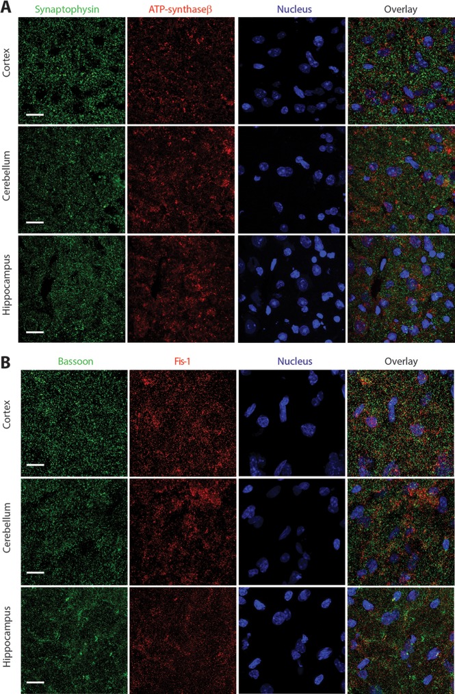
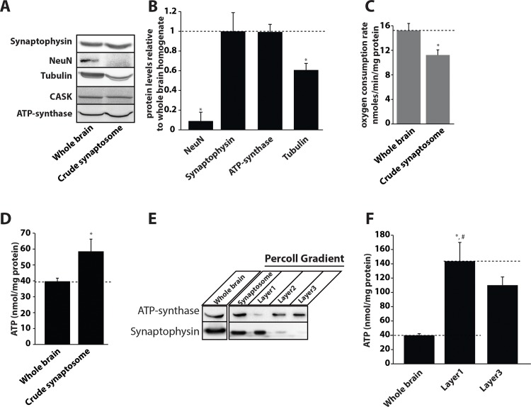
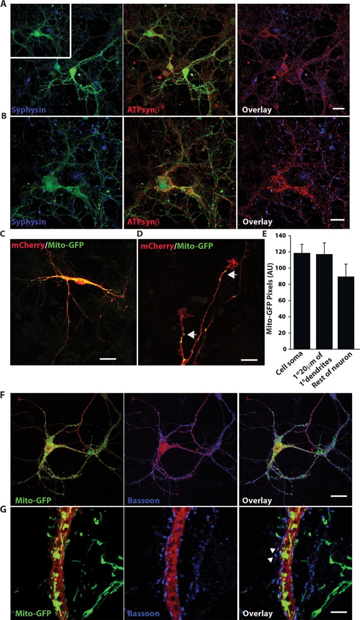
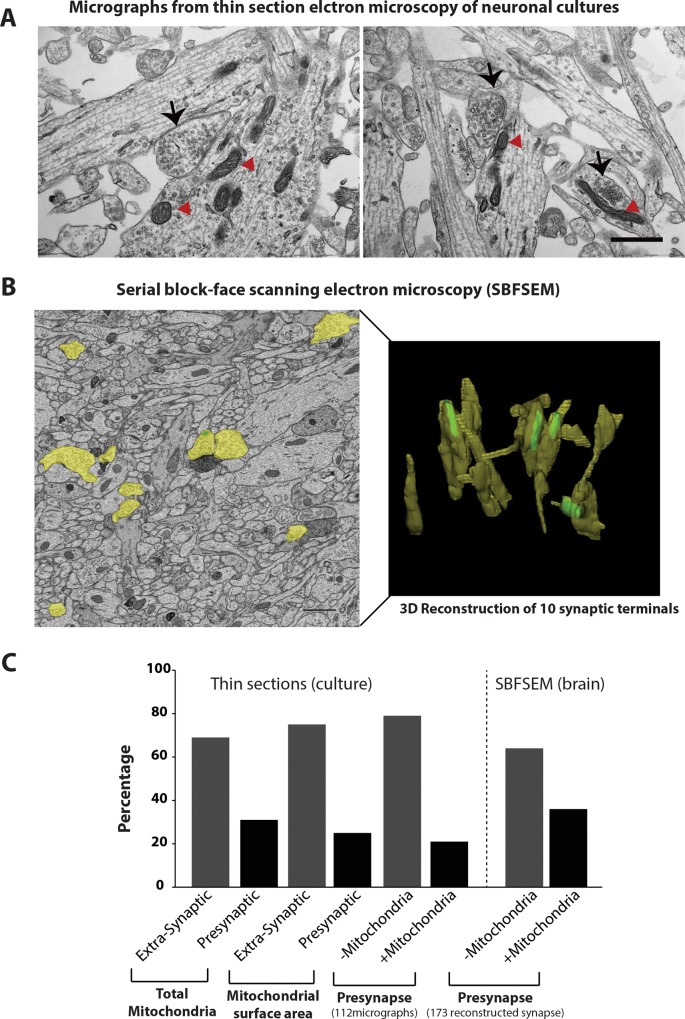
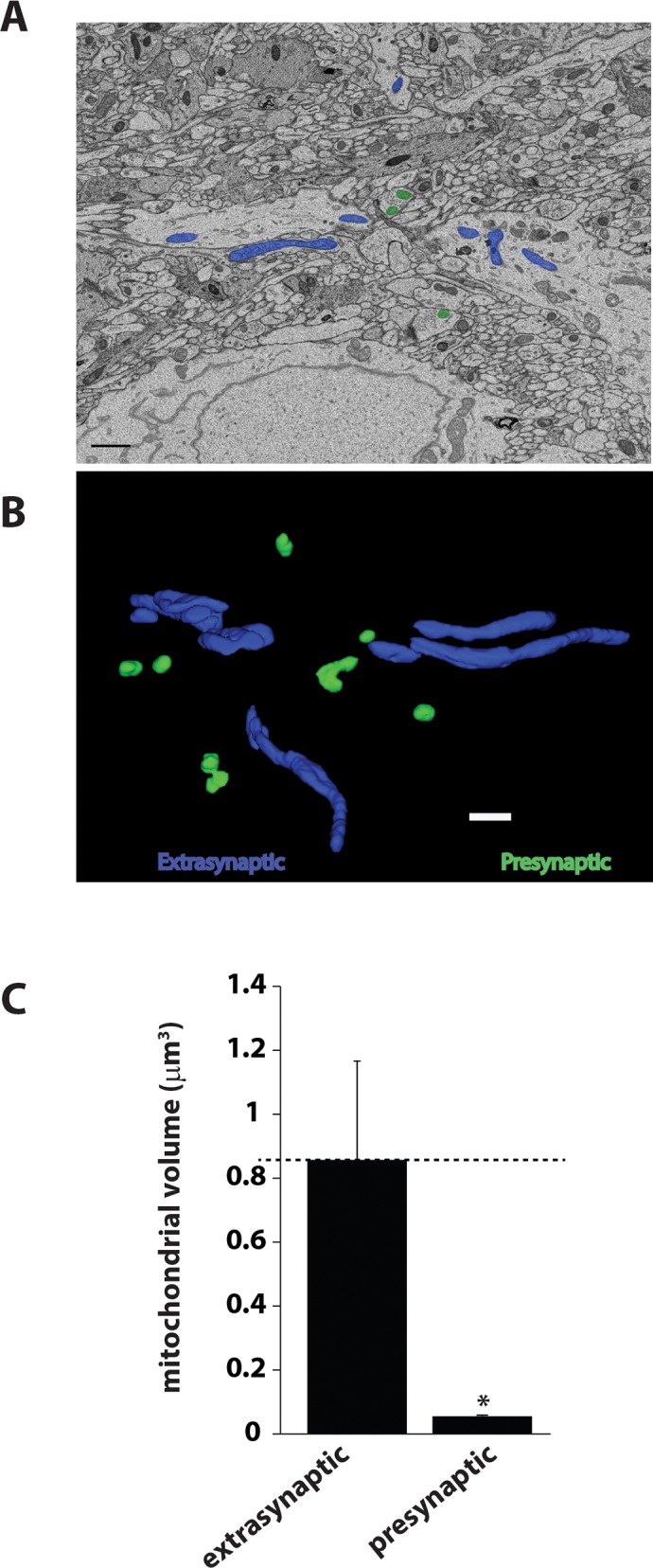
Synaptic neurotransmission is known to be an energy demanding process. At the presynapse, ATP is required for loading neurotransmitters into synaptic vesicles, for priming synaptic vesicles before release, and as a substrate for various kinases and ATPases. Although it is assumed that presynaptic sites usually harbor local mitochondria, which may serve as energy powerhouse to generate ATP as well as a presynaptic calcium depot, a clear role of presynaptic mitochondria in biochemical functioning of the presynapse is not well-defined. Besides a few synaptic subtypes like the mossy fibers and the Calyx of Held, most central presynaptic sites are either en passant or tiny axonal terminals that have little space to accommodate a large mitochondrion. Here, we have used imaging studies to demonstrate that mitochondrial antigens poorly co-localize with the synaptic vesicle clusters and active zone marker in the cerebral cortex, hippocampus and the cerebellum. Confocal imaging analysis on neuronal cultures revealed that most neuronal mitochondria are either somatic or distributed in the proximal part of major dendrites. A large number of synapses in culture are devoid of any mitochondria. Electron micrographs from neuronal cultures further confirm our finding that the majority of presynapses may not harbor resident mitochondria. We corroborated our ultrastructural findings using serial block face scanning electron microscopy (SBFSEM) and found that more than 60% of the presynaptic terminals lacked discernible mitochondria in the wild-type mice hippocampus. Biochemical fractionation of crude synaptosomes into mitochondria and pure synaptosomes also revealed a sparse presence of mitochondrial antigen at the presynaptic boutons. Despite a low abundance of mitochondria, the synaptosomal membranes were found to be highly enriched in ATP suggesting that the presynapse may possess alternative mechanism/s for concentrating ATP for its function. The potential mechanisms including local glycolysis and the possible roles of ATP-binding synaptic proteins such as synapsins, are discussed.
已知突触神经传递是一个能量需求过程。在突触前,将神经递质装载到突触小泡中、在释放前对突触小泡进行预处理以及作为各种激酶和ATP酶的底物都需要ATP。尽管一般认为突触前位点通常含有局部线粒体,其可作为产生ATP的能量源以及突触前钙库,但突触前线粒体在突触前生化功能中的明确作用尚未明确界定。除了少数突触亚型,如苔藓纤维和Held壶腹,大多数中枢突触前位点要么是过路型,要么是微小的轴突终末,几乎没有空间容纳大的线粒体。在此,我们通过成像研究证明,在大脑皮层、海马体和小脑中,线粒体抗原与突触小泡簇和活性区标记物的共定位较差。对神经元培养物的共聚焦成像分析显示,大多数神经元线粒体要么位于胞体,要么分布在主要树突的近端部分。培养物中的大量突触没有任何线粒体。神经元培养物的电子显微镜照片进一步证实了我们的发现,即大多数突触前位点可能没有驻留线粒体。我们使用连续块面扫描电子显微镜(SBFSEM)证实了我们的超微结构发现,发现在野生型小鼠海马体中,超过60%的突触前终末没有可识别的线粒体。将粗制突触体进行生物化学分级分离为线粒体和纯突触体,也显示突触前小体中线粒体抗原的存在稀少。尽管线粒体丰度较低,但发现突触体膜中ATP高度富集,这表明突触前可能具有替代机制来浓缩ATP以实现其功能。文中讨论了潜在机制,包括局部糖酵解以及诸如突触结合蛋白等ATP结合突触蛋白的可能作用。