Schmidt Martin J, Laubner Steffi, Kolecka Malgorzata, Failing Klaus, Moritz Andreas, Kramer Martin, Ondreka Nele
Department of Veterinary Clinical Sciences, Clinic for Small Animals, Justus-Liebig-University-Giessen, Giessen, Germany.
Unit for Biomathematics and Data Processing, Faculty of Veterinary Medicine, Justus Liebig-University-Giessen, Giessen, Germany.
PLoS One. 2015 May 4;10(5):e0124174. doi: 10.1371/journal.pone.0124174. eCollection 2015.
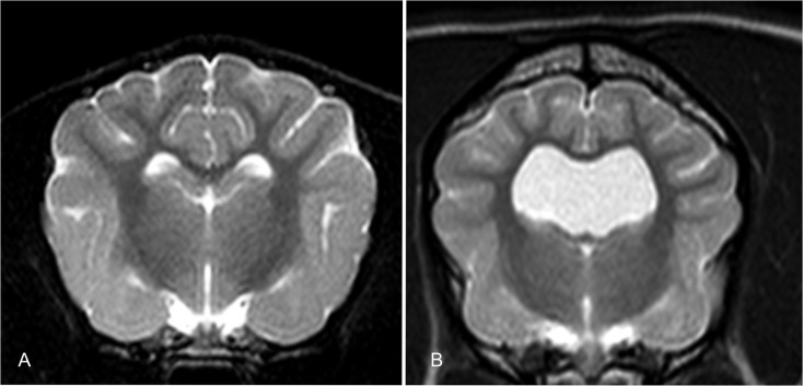
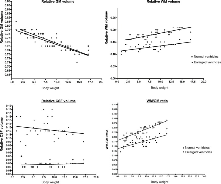
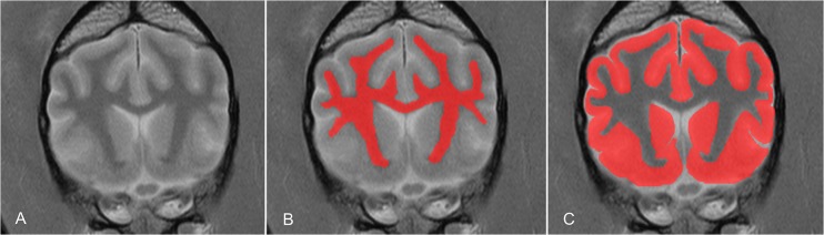
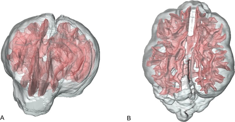
Large cerebral ventricles are a frequent finding in brains of dogs with brachycephalic skull conformation, in comparison with mesaticephalic dogs. It remains unclear whether oversized ventricles represent a normal variant or a pathological condition in brachycephalic dogs. There is a distinct relationship between white matter and grey matter in the cerebrum of all eutherian mammals. The aim of this study was to determine if this physiological proportion between white matter and grey matter of the forebrain still exists in brachycephalic dogs with oversized ventricles. The relative cerebral grey matter, white matter and cerebrospinal fluid volume in dogs were determined based on magnetic-resonance-imaging datasets using graphical software. In an analysis of covariance (ANCOVA) using body mass as the covariate, the adjusted means of the brain tissue volumes of two groups of dogs were compared. Group 1 included 37 mesaticephalic dogs of different sizes with no apparent changes in brain morphology, and subjectively normal ventricle size. Group 2 included 35 brachycephalic dogs in which subjectively enlarged cerebral ventricles were noted as an incidental finding in their magnetic-resonance-imaging examination. Whereas no significant different adjusted means of the grey matter could be determined, the group of brachycephalic dogs had significantly larger adjusted means of lateral cerebral ventricles and significantly less adjusted means of relative white matter volume. This indicates that brachycephalic dogs with subjective ventriculomegaly have less white matter, as expected based on their body weight and cerebral volume. Our study suggests that ventriculomegaly in brachycephalic dogs is not a normal variant of ventricular volume. Based on the changes in the relative proportion of WM and CSF volume, and the unchanged GM proportions in dogs with ventriculomegaly, we rather suggest that distension of the lateral ventricles might be the underlying cause of pressure related periventricular loss of white matter tissue, as occurs in internal hydrocephalus.
与中脑型犬相比,大脑脑室扩大在短头型颅骨结构犬的大脑中较为常见。目前尚不清楚在短头型犬中,过大的脑室是正常变异还是病理状况。在所有真兽类哺乳动物的大脑中,白质和灰质之间存在明显的关系。本研究的目的是确定在脑室过大的短头型犬中,前脑白质和灰质之间的这种生理比例是否仍然存在。基于磁共振成像数据集,使用图形软件确定犬的相对脑灰质、白质和脑脊液体积。在以体重作为协变量的协方差分析(ANCOVA)中,比较了两组犬脑组织体积的调整均值。第1组包括37只不同大小的中脑型犬,脑形态无明显变化,脑室大小主观上正常。第2组包括35只短头型犬,在其磁共振成像检查中偶然发现脑室主观扩大。虽然无法确定灰质的调整均值有显著差异,但短头型犬组的侧脑室调整均值显著更大,相对白质体积的调整均值显著更小。这表明,脑室主观扩大的短头型犬白质较少,这与基于它们的体重和脑体积预期的情况一致。我们的研究表明,短头型犬的脑室扩大不是脑室体积的正常变异。基于脑室扩大犬中白质和脑脊液体积相对比例的变化以及灰质比例不变的情况,我们更倾向于认为侧脑室扩张可能是脑室周围白质组织压力相关损失的潜在原因,就像在内部脑积水时发生的那样。