Liu Zhenjie, Morgan Stephanie, Ren Jun, Wang Qiwei, Annis Douglas S, Mosher Deane F, Zhang Jing, Sorenson Christine M, Sheibani Nader, Liu Bo
From the Departments of Surgery (Z.L., S.M., J.R., Q.W., B.L.), Pathology and Laboratory Medicine (B.L.), Biomolecular Chemistry and Medicine (D.S.A., D.F.M.), McArdle Laboratory for Cancer Research (J.Z.), Pediatrics (C.M.S.), and Ophthalmology and Visual Sciences (N.S.), University of Wisconsin School of Medicine and Public Health, Madison; and Department of Vascular Surgery, The Second Affiliated Hospital of Zhejiang University School of Medicine, Hangzhou, China (Z.L.).
Circ Res. 2015 Jul 3;117(2):129-41. doi: 10.1161/CIRCRESAHA.117.305262. Epub 2015 May 4.
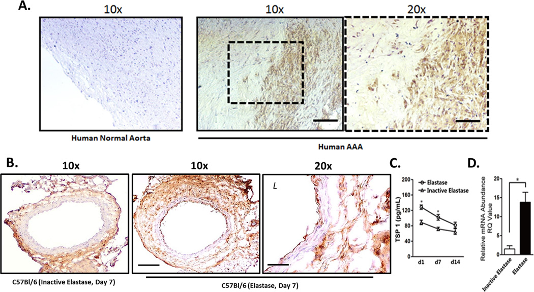
Histological examination of abdominal aortic aneurysm (AAA) tissues demonstrates extracellular matrix destruction and infiltration of inflammatory cells. Previous work with mouse models of AAA has shown that anti-inflammatory strategies can effectively attenuate aneurysm formation. Thrombospondin-1 is a matricellular protein involved in the maintenance of vascular structure and homeostasis through the regulation of biological functions, such as cell proliferation, apoptosis, and adhesion. Expression levels of thrombospondin-1 correlate with vascular disease conditions.
To use thrombospondin-1-deficient (Thbs1(-/-)) mice to test the hypothesis that thrombospondin-1 contributes to pathogenesis of AAAs.
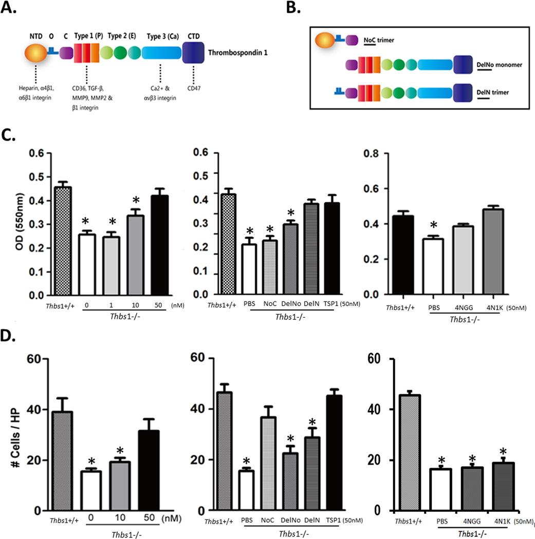
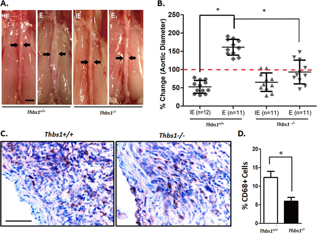
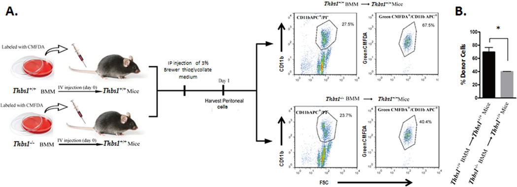
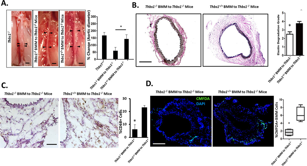
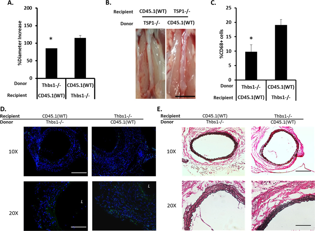
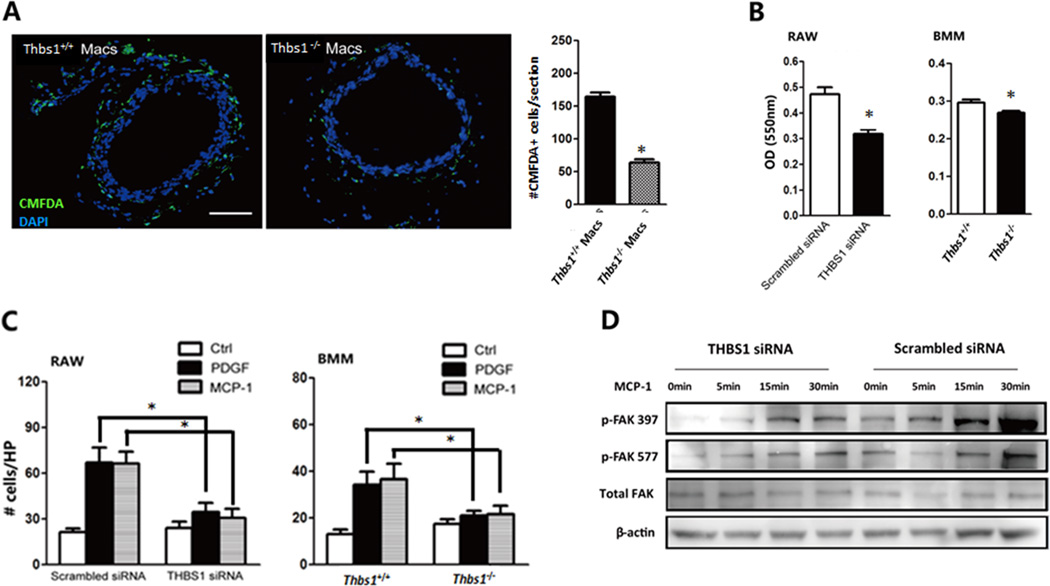
Mouse experimental AAA was induced through perivascular treatment with calcium phosphate, intraluminal perfusion with porcine elastase, or systemic administration of angiotensin II. Induction of AAA increased thrombospondin-1 expression in aortas of C57BL/6 or apoE-/- mice. Compared with Thbs1(+/+) mice, Thbs1(-/-) mice developed significantly smaller aortic expansion when subjected to AAA inductions, which was associated with diminished infiltration of macrophages. Thbs1(-/-) monocytic cells had reduced adhesion and migratory capacity in vitro compared with wild-type counterparts. Adoptive transfer of Thbs1(+/+) monocytic cells or bone marrow reconstitution rescued aneurysm development in Thbs1(-/-) mice.
Thrombospondin-1 expression plays a significant role in regulation of migration and adhesion of mononuclear cells, contributing to vascular inflammation during AAA development.
腹主动脉瘤(AAA)组织的组织学检查显示细胞外基质破坏和炎性细胞浸润。先前对AAA小鼠模型的研究表明,抗炎策略可有效减轻动脉瘤形成。血小板反应蛋白-1是一种基质细胞蛋白,通过调节细胞增殖、凋亡和黏附等生物学功能参与维持血管结构和稳态。血小板反应蛋白-1的表达水平与血管疾病状况相关。
利用血小板反应蛋白-1缺陷(Thbs1(-/-))小鼠来验证血小板反应蛋白-1促成AAA发病机制的假说。
通过血管周围注射磷酸钙、腔内灌注猪弹性蛋白酶或全身给予血管紧张素II诱导小鼠实验性AAA。AAA诱导增加了C57BL/6或载脂蛋白E基因敲除(apoE-/-)小鼠主动脉中血小板反应蛋白-1的表达。与Thbs1(+/+)小鼠相比,Thbs1(-/-)小鼠在接受AAA诱导时主动脉扩张明显较小,这与巨噬细胞浸润减少有关。与野生型单核细胞相比,Thbs1(-/-)单核细胞在体外的黏附和迁移能力降低。过继转移Thbs1(+/+)单核细胞或骨髓重建可挽救Thbs1(-/-)小鼠的动脉瘤发展。
血小板反应蛋白-1的表达在调节单核细胞的迁移和黏附中起重要作用,在AAA发展过程中促成血管炎症。