Tripathy Kalpalata, Misra Aparajita, Ghosh Joydip Kumar
Department of Pathology, Shrirama Chandra Bhanja Medical College, Cuttack, Odisha, India.
J Cytol. 2015 Jan-Mar;32(1):17-20. doi: 10.4103/0970-9371.155225.
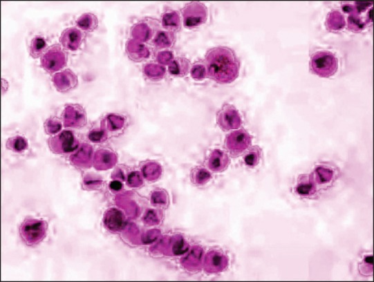
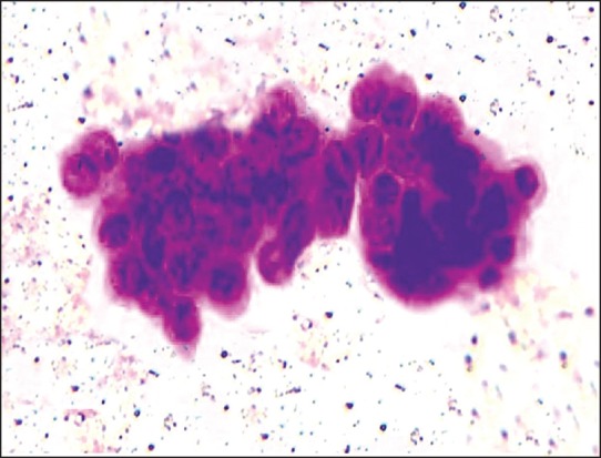
Liquid-based cytology (LBC) is fast becoming a useful method in evaluating both gynecological and non-gynecological preparations, including fine needle aspiration (FNA) cytology. Even distribution of cells, decreasing obscuring background elements like blood and mucus, well preserved nuclear and cytoplasmic details and rapid fixation helps in better visualization of cells.
This study was conducted to asses the diagnostic accuracy of liquid-based cytology versus conventional smears in fine needle aspiration samples.
In this prospective study, we had 110 cases, including 30 cases of breast, 40 of lymph node, 10 of salivary glands, 18 of thyroid and 12 of bone and soft tissue. In each case, two passes were performed. The first pass was for conventional preparation (CP) and the second pass yielded material for thin-prep (TP) preparation. Both CP and TP smears were compared for cellularity, background blood and necrotic cell debris, cell architecture, informative background, presence of a monolayer of cells and nuclear and cytoplasmic details by a semiquantitative scoring system. Wilcoxon's signed rank test on an SPSS program was used for statistical analysis.
Diagnostic accuracy was better in LBC smears compared with CP smears due to lack of background debris and better cell morphology, which was performed according to Wilcoxon's signed rank test, yielding a P-value of <0.001. However, in some cases, because of a decrease in cell size, clustering and altered background in LBC, a support of CP was essential.
LBC performed on FNA samples can be a simple and valuable technique. Only in few selected cases, where background factor is an essential diagnostic clue, a combination of both CP and TP is necessary.
液基细胞学(LBC)正迅速成为评估妇科和非妇科标本(包括细针穿刺(FNA)细胞学)的一种有用方法。细胞分布均匀、减少血液和黏液等模糊背景成分、细胞核和细胞质细节保存良好以及快速固定有助于更好地观察细胞。
本研究旨在评估液基细胞学与传统涂片在细针穿刺样本中的诊断准确性。
在这项前瞻性研究中,我们纳入了110例病例,包括30例乳腺病例、40例淋巴结病例、10例唾液腺病例、18例甲状腺病例以及12例骨和软组织病例。对每个病例进行两次穿刺。第一次穿刺用于传统制片(CP),第二次穿刺获取的材料用于薄层制片(TP)。通过半定量评分系统对CP和TP涂片的细胞数量、背景血液和坏死细胞碎片、细胞结构、信息背景、单层细胞的存在情况以及细胞核和细胞质细节进行比较。使用SPSS软件中的Wilcoxon符号秩检验进行统计分析。
根据Wilcoxon符号秩检验,由于缺乏背景碎片且细胞形态更好,LBC涂片的诊断准确性优于CP涂片,P值<0.001。然而,在某些情况下,由于LBC中细胞大小减小、细胞聚集以及背景改变,CP的支持是必不可少的。
对FNA样本进行LBC检查可以是一种简单且有价值的技术。仅在少数选定的病例中,当背景因素是关键诊断线索时,需要将CP和TP联合使用。