Neurology Department, University of Melbourne Veterinary Clinic and Hospital, 250 Princes Highway, Werribee, Melbourne, 3030 Australia.
Internal Medicine Department, University of Melbourne Veterinary Clinic and Hospital, 250 Princes Highway, Werribee, Melbourne, 3030 Australia.
Ir Vet J. 2015 Apr 24;68(1):5. doi: 10.1186/s13620-015-0033-6. eCollection 2015.
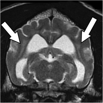
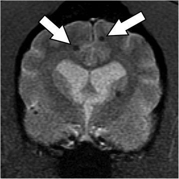
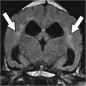
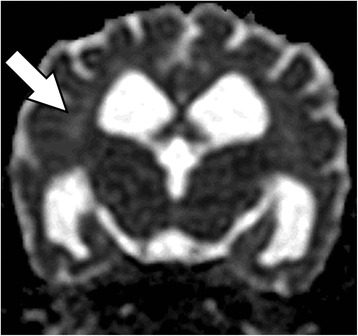
A 16-year-old female spayed English Staffordshire terrier was presented for evaluation of a 10-month history of intermittent myoclonic episodes, and a one weeks history of short episodes of altered mentation, ataxia and collapse. Magnetic resonance imaging identified subcortical oedema, predominately in the parietal and temporal lobes and multiple cerebral microbleeds. Serum biochemistry, indirect blood pressure measurements and magnetic resonance imaging abnormalities were consistent with hypertensive encephalopathy secondary to chronic kidney disease.
一只 16 岁已绝育的雌性英国史宾格犬,因间歇性肌阵挛发作 10 个月,以及精神状态改变、共济失调和瘫痪的短暂发作 1 周就诊。磁共振成像发现皮质下水肿,主要位于顶叶和颞叶,并有多处脑微出血。血清生化、间接血压测量和磁共振成像异常与慢性肾病继发的高血压性脑病一致。