Miguel-Garcés María, Gonçalves Rita, Quintana Rodrigo, Álvarez Patricia, Beckmann Katrin M, Alcoverro Emili, Moioli Melania, Ives Edward J, Madden Megan, Gomes Sergio A, Galban Evelyn, Bentley Tim, Santifort Koen M, Vanhaesebrouck An, Briola Chiara, Montoliu Patricia, Ibaseta Unai, Carrera Inés
Diagnostic Imaging Department, Southern Counties Veterinary Specialists, Independent Vetcare (IVC) Evidensia, Ringwood, United Kingdom.
Department of Small Animal Clinical Science, Small Animal Teaching Hospital, University of Liverpool, Neston, United Kingdom.
Front Vet Sci. 2024 Jul 30;11:1390971. doi: 10.3389/fvets.2024.1390971. eCollection 2024.
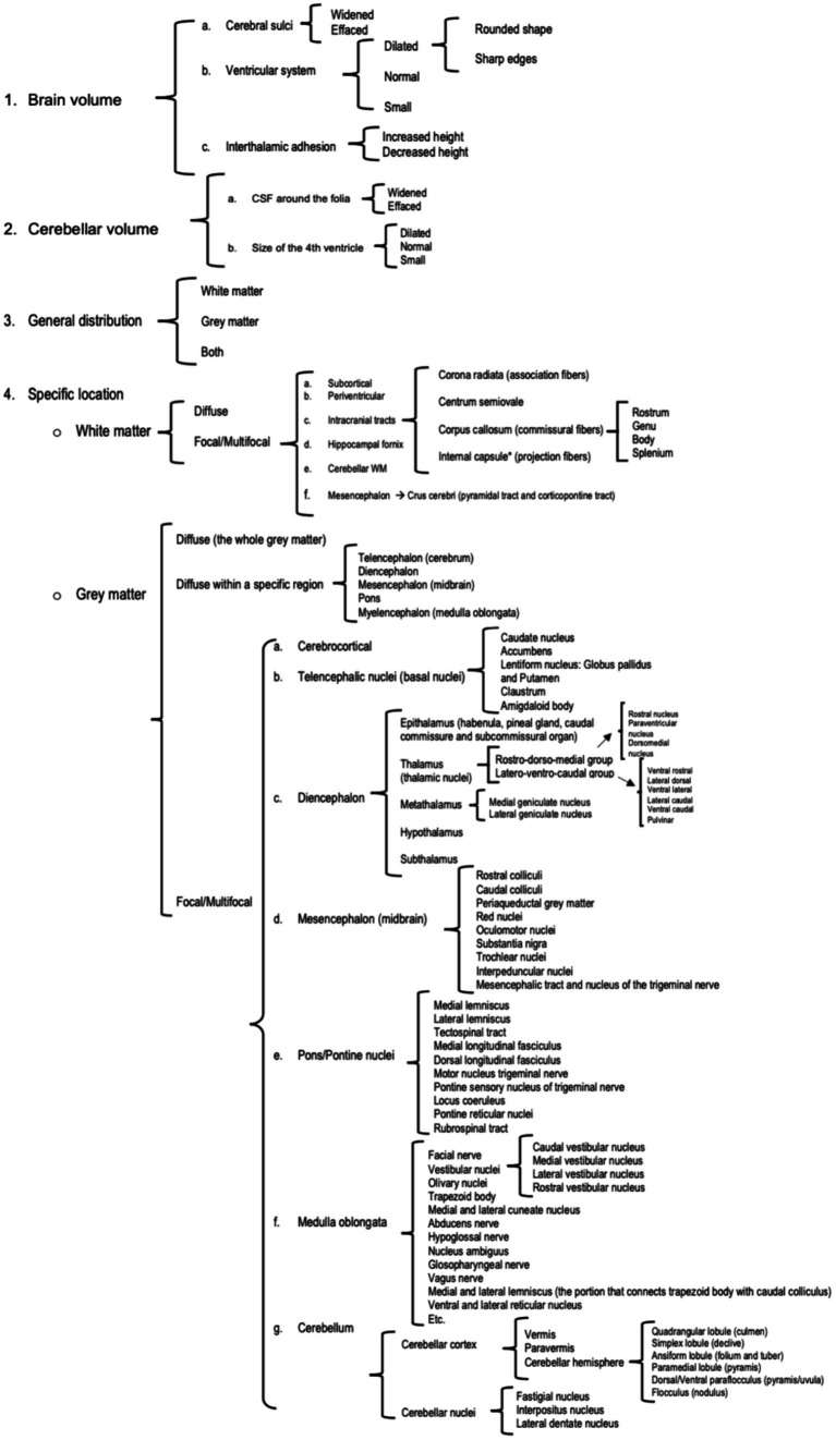
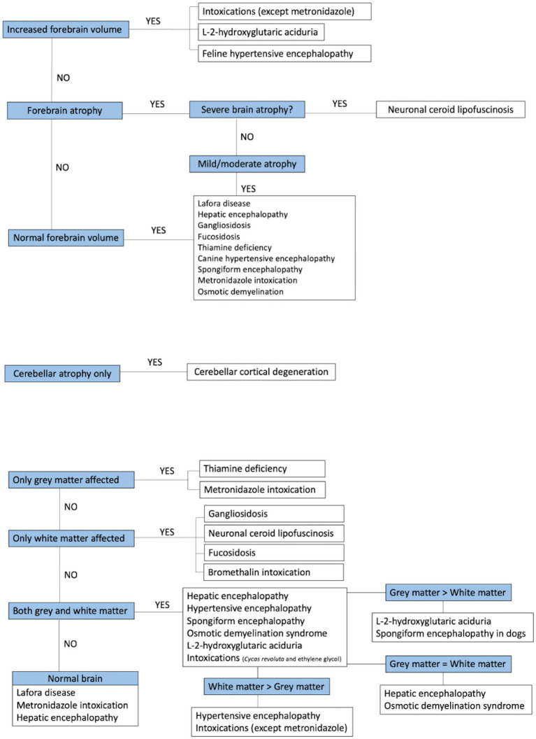
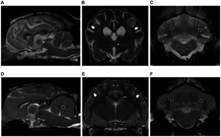
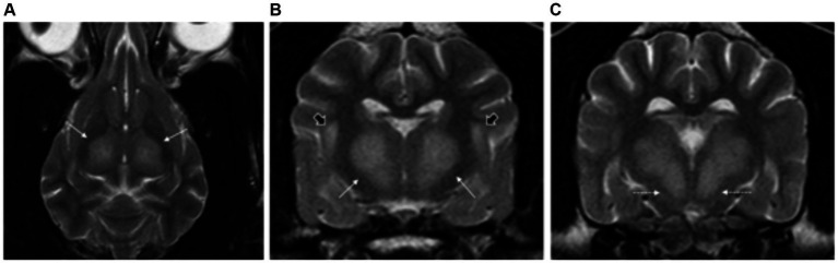
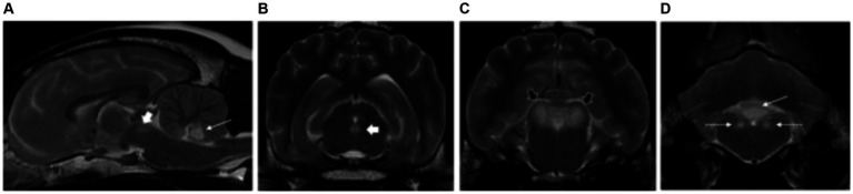
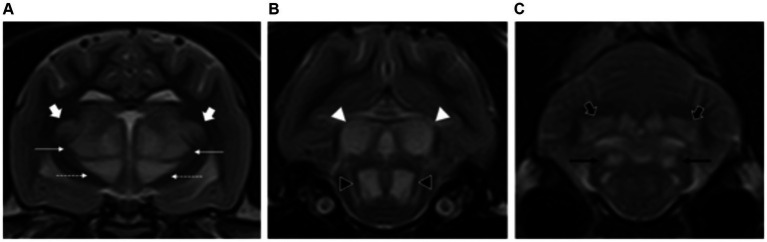
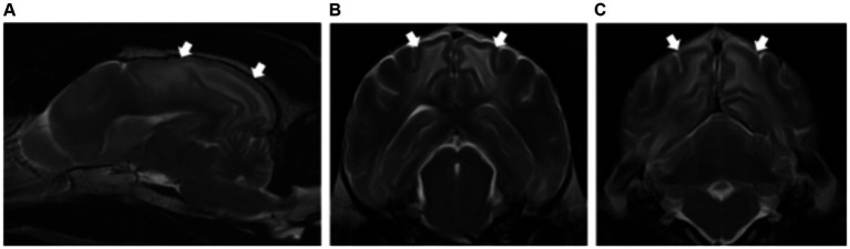
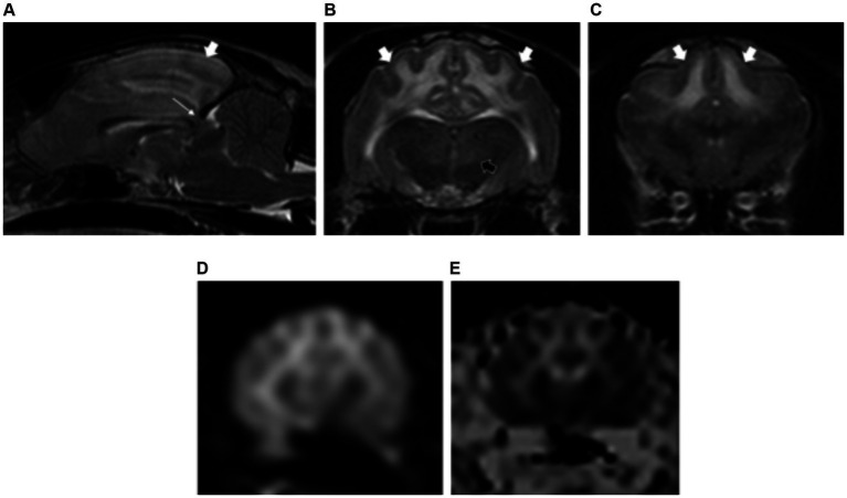
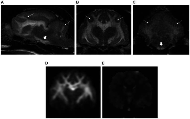
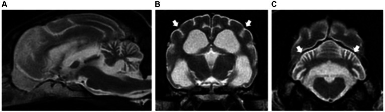
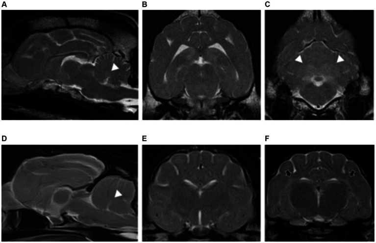
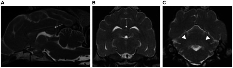
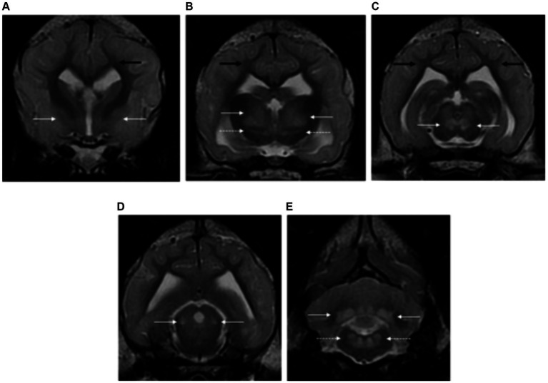
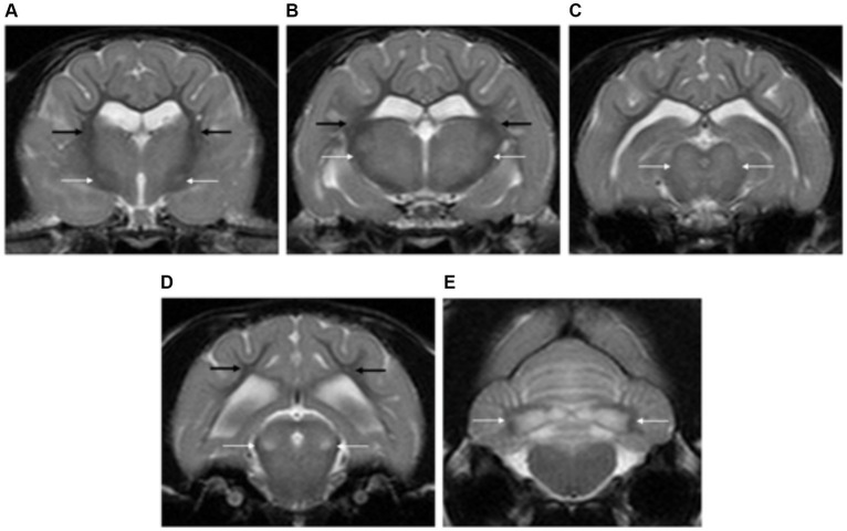
Metabolic/neurodegenerative encephalopathies encompass a wide list of conditions that share similar clinical and magnetic resonance imaging (MRI) characteristics, challenging the diagnostic process and resulting in numerous tests performed in order to reach a definitive diagnosis. The aims of this multicentric, retrospective and descriptive study are: (I) to describe the MRI features of dogs and cats with metabolic/neurodegenerative encephalopathies; (II) to attempt an MRI recognition pattern classifying these conditions according to the involvement of grey matter, white matter or both; and (III) to correlate the MRI findings with previous literature. A total of 100 cases were recruited, comprising 81 dogs and 19 cats. These included hepatic encephalopathy (20 dogs and three cats), myelinolysis (five dogs), intoxications (seven dogs and one cat), thiamine deficiency (two dogs and seven cats), hypertensive encephalopathy (three dogs and two cats), neuronal ceroid lipofuscinosis (11 dogs and one cat), gangliosidosis (three dogs and two cats), fucosidosis (one dog), L-2-hydroxyglutaric aciduria (13 dogs and one cat), Lafora disease (11 dogs), spongiform leukoencephalomyelopathy (one dog) and cerebellar cortical degeneration (four dogs and two cats). None of the hepatic encephalopathies showed the previously described T1-weighted hyperintensity of the lentiform nuclei. Instead, there was involvement of the cerebellar nuclei (8/23), which is a feature not previously described. Dogs with myelinolysis showed novel involvement of a specific white matter structure, the superior longitudinal fasciculus (5/5). Thiamine deficiency affected numerous deep grey nuclei with novel involvement of the oculomotor nuclei (3/9), thalamic nuclei, subthalamus and cerebellar nuclei (1/9). Cats with hypertensive encephalopathy had a more extensive distribution of the white matter changes when compared to dogs, extending from the parietal and occipital lobes into the frontal lobes with associated mass effect and increased brain volume. Lysosomal storage disease showed white matter involvement only, with neuronal ceroid lipofuscinosis characterised by severe brain atrophy when compared to gangliosidosis and fucosidosis. All patients with L-2-hydroxyglutaric aciduria had a characteristic T2-weighted hyperintense swelling of the cerebral and cerebellar cortical grey matter, resulting in increased brain volume. Lafora disease cases showed either normal brain morphology (5/11) or mild brain atrophy (6/11). Dogs with cerebellar cortical degeneration had more marked cerebellar atrophy when compared to cats. This study shows the important role of MRI in distinguishing different metabolic/neurodegenerative encephalopathies according to specific imaging characteristics.
代谢性/神经退行性脑病包含一系列具有相似临床和磁共振成像(MRI)特征的病症,这给诊断过程带来了挑战,导致需要进行大量检查以得出明确诊断。这项多中心、回顾性和描述性研究的目的是:(I)描述患有代谢性/神经退行性脑病的犬猫的MRI特征;(II)尝试根据灰质、白质或两者的受累情况对这些病症进行MRI识别模式分类;(III)将MRI结果与先前的文献进行关联。共招募了100例病例,其中包括81只犬和19只猫。这些病例包括肝性脑病(20只犬和3只猫)、髓鞘溶解症(5只犬)、中毒(7只犬和1只猫)、硫胺素缺乏症(2只犬和7只猫)、高血压脑病(3只犬和2只猫)、神经元蜡样脂褐质沉积症(11只犬和1只猫)、神经节苷脂贮积症(3只犬和2只猫)、岩藻糖苷贮积症(1只犬)、L-2-羟基戊二酸尿症(13只犬和1只猫)、拉福拉病(11只犬)、海绵状白质脑病(1只犬)和小脑皮质变性(4只犬和2只猫)。所有肝性脑病病例均未显示出先前描述的豆状核T1加权高信号。相反,小脑核受累(8/23),这是一个先前未描述的特征。患有髓鞘溶解症的犬出现了一种特定白质结构——上纵束的新受累情况(5/5)。硫胺素缺乏症影响了许多深部灰质核,动眼神经核(3/9)、丘脑核、下丘脑和小脑核出现了新的受累情况(1/9)。与犬相比,患有高血压脑病的猫白质变化分布更广泛,从顶叶和枕叶延伸至额叶,伴有相关的占位效应和脑容量增加。溶酶体贮积症仅表现为白质受累,与神经节苷脂贮积症和岩藻糖苷贮积症相比,神经元蜡样脂褐质沉积症的特征是严重脑萎缩。所有L-2-羟基戊二酸尿症患者的大脑和小脑皮质灰质在T2加权上均有特征性的高信号肿胀,导致脑容量增加。拉福拉病病例的脑形态要么正常(5/11),要么轻度脑萎缩(6/11)。与猫相比,患有小脑皮质变性的犬小脑萎缩更明显。这项研究表明MRI在根据特定成像特征区分不同的代谢性/神经退行性脑病方面具有重要作用。