Dagenais Marie, MacDonald David, Baron Murray, Hudson Marie, Tatibouet Solène, Steele Russell, Gravel Sabrina, Mohit Shrisha, El Sayegh Tarek, Pope Janet, Fontaine Audrey, Masseto Ariel, Matthews Debora, Sutton Evelyn, Thie Norman, Jones Niall, Copete Maria, Kolbinson Dean, Markland Janet, Nogueira-Filho Getulio, Robinson David, Gornitsky Mervyn
Faculty of Dentistry, McGill University, 2001 McGill College Avenue, Suite 500, Montreal, Quebec H3A 1G1.
Faculty of Dentistry, Division of Oral & Maxillofacial Radiology, University of British Columbia, Room 380, J.B. Macdonald Building, 2199 Wesbrook Mall, Vancouver, British Columbia V6T 1Z3.
Oral Surg Oral Med Oral Pathol Oral Radiol. 2015 Aug;120(2):104-11. doi: 10.1016/j.oooo.2015.03.002. Epub 2015 Mar 25.
The aim of this study was to compare oral radiologic abnormalities associated with systemic sclerosis (SSc) against abnormalities in the general population.
Patients with SSc and healthy controls were enrolled in a multi-site cross-sectional study. Included in the radiology examination were a panoramic radiograph, four bitewings, and an anterior mandibular periapical radiograph. Radiographs were evaluated by two oral and maxillofacial radiologists tested for interobserver and intraobserver reliability. Chi-squared tests, Fisher exact tests, and Mann Whitney U tests were used to summarize the radiologic manifestations of patients and controls.
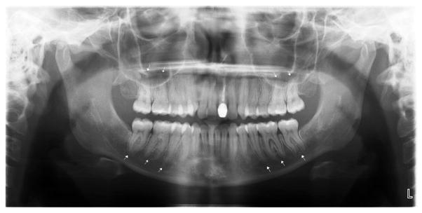
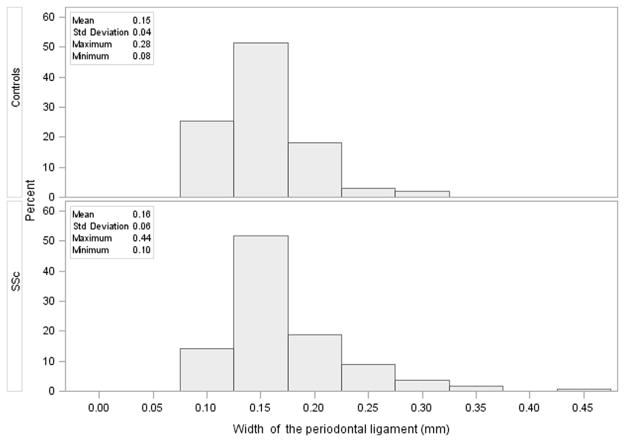
We assessed 163 SSc patients and 231 controls. Widening of the periodontal ligament space (PLS) (P < .001), with higher percentage of teeth with PLS widening (P < .001), was significantly more frequent in patients with SSc than in controls. The most significant differences between the two groups were found in the molars and premolars (P < .001). Moreover, 26% of the patients with SSc had a periapical PLS greater than 0.19 mm compared with 13% of the controls (P = .003). Patients with SSc had significantly more erosions compared with controls (14.5% vs. 3.6%; P < .001), mostly in the condyles (P = .022), coronoid processes (P = .005) and other locations (P = .012).
Patients with SSc had more teeth with PLS widening and erosions of the mandible compared with controls.
本研究旨在比较系统性硬化症(SSc)患者口腔放射学异常与普通人群异常情况。
SSc患者和健康对照者纳入一项多中心横断面研究。放射学检查包括一张全景X线片、四张咬合翼片和一张下颌前部根尖片。由两名口腔颌面放射科医生对X线片进行评估,并检测观察者间和观察者内的可靠性。采用卡方检验、Fisher精确检验和Mann-Whitney U检验总结患者和对照者的放射学表现。
我们评估了163例SSc患者和231例对照者。SSc患者牙周膜间隙(PLS)增宽(P <.001),PLS增宽牙齿的百分比更高(P <.001),显著高于对照者。两组之间最显著的差异见于磨牙和前磨牙(P <.001)。此外,26%的SSc患者根尖PLS大于0.19 mm,而对照者为13%(P =.003)。与对照者相比,SSc患者的侵蚀明显更多(14.5%对3.6%;P <.001),主要见于髁突(P =.022)、冠状突(P =.005)和其他部位(P =.012)。
与对照者相比,SSc患者PLS增宽的牙齿更多,下颌骨侵蚀更多。