Strobel Claudia, Oehring Hartmut, Herrmann Rudolf, Förster Martin, Reller Armin, Hilger Ingrid
Department of Experimental Radiology, Institute of Diagnostic and Interventional Radiology, Jena University Hospital - Friedrich Schiller University Jena, Erlanger Allee 101, 07747 Jena, Germany.
Institute of Anatomy II, Jena University Hospital - Friedrich Schiller University Jena, Teichgraben 7, 07743 Jena, Germany.
J Nanopart Res. 2015;17(5):206. doi: 10.1007/s11051-015-3007-4. Epub 2015 May 5.
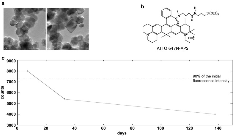
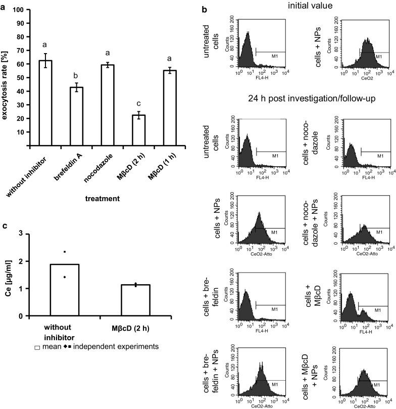
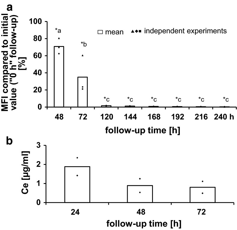
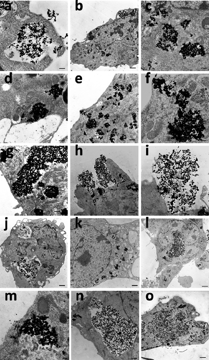
Although cytotoxicity and endocytosis of nanoparticles have been the subject of numerous studies, investigations regarding exocytosis as an important mechanism to reduce intracellular nanoparticle accumulation are rather rare and there is a distinct lack of knowledge. The current study investigated the behavior of human microvascular endothelial cells to exocytose cerium dioxide (CeO) nanoparticles (18.8 nm) by utilization of specific inhibitors [brefeldin A; nocodazole; methyl-β-cyclodextrin (MβcD)] and different analytical methods (flow cytometry, transmission electron microscopy, inductively coupled plasma mass spectrometry). Overall, it was found that endothelial cells were able to release CeO nanoparticles via exocytosis after the migration of nanoparticle containing endosomes toward the plasma membrane. The exocytosis process occurred mainly by fusion of vesicular membranes with plasma membrane resulting in the discharge of vesicular content to extracellular environment. Nevertheless, it seems to be likely that nanoparticles present in the cytosol could leave the cells in a direct manner. MβcD treatment led to the strongest inhibition of the nanoparticle exocytosis indicating a significant role of the plasma membrane cholesterol content in the exocytosis process. Brefeldin A (inhibitor of Golgi-to-cell-surface-transport) caused a higher inhibitory effect on exocytosis than nocodazole (inhibitor of microtubules). Thus, the transfer from distal Golgi compartments to the cell surface influenced the exocytosis process of the CeO nanoparticles more than the microtubule-associated transport. In conclusion, endothelial cells, which came in contact with nanoparticles, e.g., after intravenously applied nano-based drugs, can regulate their intracellular nanoparticle amount, which is necessary to avoid adverse nanoparticle effects on cells.
尽管纳米颗粒的细胞毒性和内吞作用已成为众多研究的主题,但关于胞吐作用作为减少细胞内纳米颗粒积累的重要机制的研究却相当少见,且明显缺乏相关知识。本研究通过使用特定抑制剂[布雷菲德菌素A;诺考达唑;甲基-β-环糊精(MβcD)]和不同分析方法(流式细胞术、透射电子显微镜、电感耦合等离子体质谱),研究了人微血管内皮细胞对二氧化铈(CeO)纳米颗粒(18.8纳米)的胞吐行为。总体而言,发现内皮细胞在含纳米颗粒的内体向质膜迁移后,能够通过胞吐作用释放CeO纳米颗粒。胞吐过程主要通过囊泡膜与质膜融合,导致囊泡内容物排放到细胞外环境。然而,似乎胞质溶胶中存在的纳米颗粒可能直接离开细胞。MβcD处理对纳米颗粒胞吐作用的抑制作用最强,表明质膜胆固醇含量在胞吐过程中起重要作用。布雷菲德菌素A(高尔基体到细胞表面运输的抑制剂)对胞吐作用的抑制作用比诺考达唑(微管抑制剂)更高。因此,从高尔基体远端区室到细胞表面的转运对CeO纳米颗粒胞吐过程的影响大于微管相关运输。总之,与纳米颗粒接触的内皮细胞,例如在静脉内应用纳米药物后,能够调节其细胞内纳米颗粒的数量,这对于避免纳米颗粒对细胞产生不良影响是必要的。