Yassen Ghaeth Hamdon, Eckert George Joseph, Platt Jeffrey Allen
Department of Restorative Dentistry, Division of Dental Biomaterials, Indiana University School of Dentistry, Indianapolis, IN, USA.
Department of Biostatistics, Indiana University School of Medicine, Indianapolis, IN, USA.
Restor Dent Endod. 2015 May;40(2):104-12. doi: 10.5395/rde.2015.40.2.104. Epub 2014 Dec 24.
This study was performed to investigate the effects of different intracanal medicaments on chemical structure and microhardness of dentin.
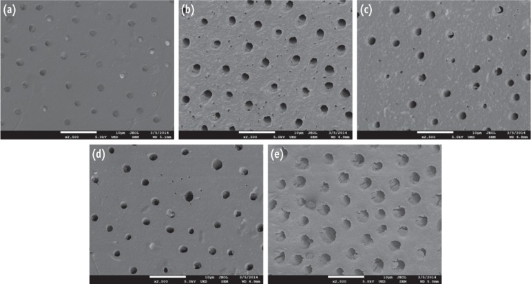
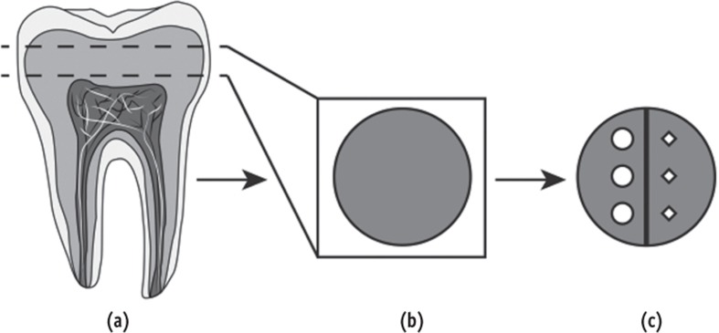
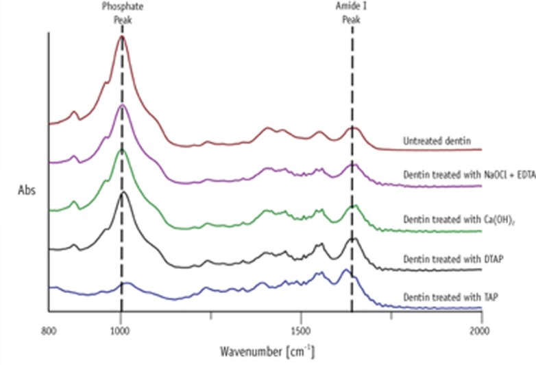
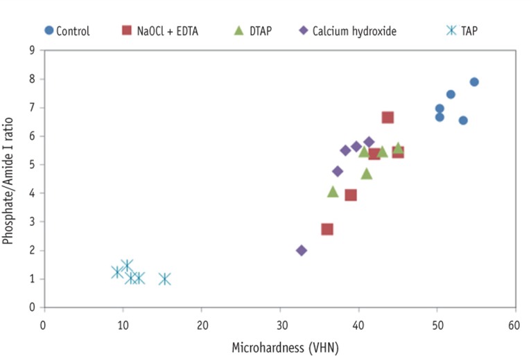
Fifty human dentin discs were obtained from intact third molars and randomly assigned into two control groups and three treatment groups. The first control group received no treatment. The second control group (no medicament group) was irrigated with sodium hypochlorite (NaOCl), stored in humid environment for four weeks and then irrigated with ethylenediaminetetraacetic acid (EDTA). The three treatment groups were irrigated with NaOCl, treated for four weeks with either 1 g/mL triple antibiotic paste (TAP), 1 mg/mL methylcellulose-based triple antibiotic paste (DTAP), or calcium hydroxide [Ca(OH)2] and finally irrigated with EDTA. After treatment, one half of each dentin disc was subjected to Vickers microhardness (n = 10 per group) and the other half was used to evaluate the chemical structure (phosphate/amide I ratio) of treated dentin utilizing attenuated total reflection Fourier transform infrared spectroscopy (n = 5 per group). One-way ANOVA followed by Fisher's least significant difference were used for statistical analyses.
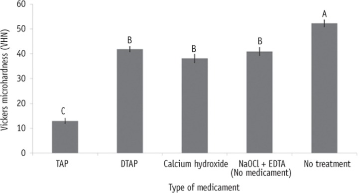
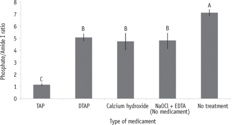
Dentin discs treated with different intracanal medicaments and those treated with NaOCl + EDTA showed significant reduction in microhardness (p < 0.0001) and phosphate/amide I ratio (p < 0.05) compared to no treatment control dentin. Furthermore, dentin discs treated with TAP had significantly lower microhardness (p < 0.0001) and phosphate/amide I ratio (p < 0.0001) compared to all other groups.
The use of DTAP or Ca(OH)2 medicaments during endodontic regeneration may cause significantly less microhardness reduction and superficial demineralization of dentin compared to the use of TAP.
本研究旨在探讨不同根管内药物对牙本质化学结构和显微硬度的影响。
从完整的第三磨牙获取50个人类牙本质盘,并随机分为两个对照组和三个治疗组。第一个对照组不进行处理。第二个对照组(无药物组)用次氯酸钠(NaOCl)冲洗,在潮湿环境中储存四周,然后用乙二胺四乙酸(EDTA)冲洗。三个治疗组用NaOCl冲洗,分别用1 g/mL三联抗生素糊剂(TAP)、1 mg/mL甲基纤维素基三联抗生素糊剂(DTAP)或氢氧化钙[Ca(OH)2]处理四周,最后用EDTA冲洗。处理后,每个牙本质盘的一半进行维氏显微硬度测试(每组n = 10),另一半用于利用衰减全反射傅里叶变换红外光谱评估处理后牙本质的化学结构(磷酸盐/酰胺I比率)(每组n = 5)。采用单因素方差分析和Fisher最小显著差法进行统计分析。
与未处理的对照牙本质相比,用不同根管内药物处理的牙本质盘以及用NaOCl + EDTA处理的牙本质盘的显微硬度(p < 0.0001)和磷酸盐/酰胺I比率(p < 0.05)显著降低。此外,与所有其他组相比,用TAP处理的牙本质盘的显微硬度(p < 0.0001)和磷酸盐/酰胺I比率(p < 0.0001)显著更低。
与使用TAP相比,在牙髓再生过程中使用DTAP或Ca(OH)2药物可能导致牙本质的显微硬度降低和表面脱矿显著减少。