• 文献检索
  • 文档翻译
  • 深度研究
  • 学术资讯
  • Suppr Zotero 插件Zotero 插件
  • 邀请有礼
  • 套餐&价格
  • 历史记录
应用&插件
Suppr Zotero 插件Zotero 插件浏览器插件Mac 客户端Windows 客户端微信小程序
定价
高级版会员购买积分包购买API积分包
服务
文献检索文档翻译深度研究API 文档MCP 服务
关于我们
关于 Suppr公司介绍联系我们用户协议隐私条款
关注我们

Suppr 超能文献

核心技术专利:CN118964589B侵权必究
粤ICP备2023148730 号-1Suppr @ 2026

文献检索

告别复杂PubMed语法,用中文像聊天一样搜索,搜遍4000万医学文献。AI智能推荐,让科研检索更轻松。

立即免费搜索

文件翻译

保留排版,准确专业,支持PDF/Word/PPT等文件格式,支持 12+语言互译。

免费翻译文档

深度研究

AI帮你快速写综述,25分钟生成高质量综述,智能提取关键信息,辅助科研写作。

立即免费体验

延迟计算机断层扫描关节造影术在体内对人膝关节软骨的评估。

Delayed Computed Tomography Arthrography of Human Knee Cartilage In Vivo.

机构信息

Department of Applied Physics, University of Eastern Finland, Kuopio, Finland ; Department of Clinical Neurophysiology, Kuopio University Hospital, Kuopio, Finland.

Department of Medical Physics, Imaging Centre, Tampere University Hospital, Tampere, Finland.

出版信息

Cartilage. 2012 Oct;3(4):334-41. doi: 10.1177/1947603512447300.

DOI:10.1177/1947603512447300
PMID:26069643
原文链接:https://pmc.ncbi.nlm.nih.gov/articles/PMC4297151/
Abstract

OBJECTIVE

We investigated the feasibility of delayed computed tomography (CT) arthrography for evaluation of human knee cartilage in vivo. Especially, the diffusion of contrast agent out of the joint space and the optimal time points for imaging were determined.

DESIGN

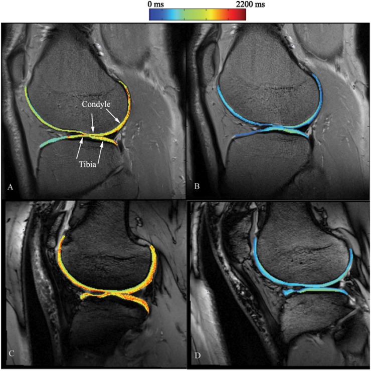
Two patients were imaged using delayed CT arthrography and delayed gadolinium-enhanced magnetic resonance imaging of cartilage (dGEMRIC) techniques.

RESULTS

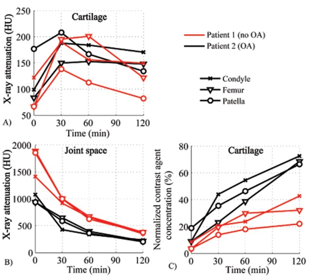
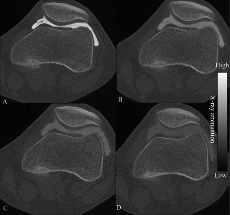
Two hours after injection, the concentration of contrast agent in the joint space was still high enough (20% to 24.5% of the initial concentration at 0 minutes) to allow delayed CT arthrography. The half-life of the contrast agent in the joint space varied from 30 to 60 minutes. The contrast agent concentration in patellar and femoral cartilage reached the maximum after 30 and 60 minutes, respectively. According to dGEMRIC, there were no differences between patients. However, in delayed CT arthrography, the penetration of the contrast agent was higher in the osteoarthritic knee cartilage.

CONCLUSIONS

Contrast agent remained in the joint space long enough to enable delayed CT arthrography of cartilage. After 30 minutes, the normalized contrast agent concentration was higher in the cartilage of the osteoarthritic knee in comparison with the healthy knee. To conclude, delayed CT arthrography exhibited potential for use in the clinical evaluation of cartilage integrity.

摘要

目的

我们研究了延迟 CT 关节造影术评估活体人膝关节软骨的可行性。特别是,确定了对比剂从关节腔扩散的情况以及成像的最佳时间点。

设计

两名患者分别使用延迟 CT 关节造影和延迟钆增强磁共振成像软骨(dGEMRIC)技术进行成像。

结果

注射后 2 小时,关节腔内的对比剂浓度仍然足够高(0 分钟时初始浓度的 20%至 24.5%),可以进行延迟 CT 关节造影。关节腔内对比剂的半衰期从 30 分钟到 60 分钟不等。髌股和股骨软骨中的对比剂浓度分别在 30 分钟和 60 分钟达到最大值。根据 dGEMRIC,患者之间没有差异。然而,在延迟 CT 关节造影中,骨关节炎膝关节软骨的对比剂渗透更高。

结论

对比剂在关节腔内停留的时间足以进行软骨的延迟 CT 关节造影。30 分钟后,与健康膝关节相比,骨关节炎膝关节软骨中的归一化对比剂浓度更高。总之,延迟 CT 关节造影术在评估软骨完整性方面具有应用潜力。

https://cdn.ncbi.nlm.nih.gov/pmc/blobs/4987/4297151/bdf15e9bf515/10.1177_1947603512447300-fig4.jpg
https://cdn.ncbi.nlm.nih.gov/pmc/blobs/4987/4297151/ef8829fd83d8/10.1177_1947603512447300-fig1.jpg
https://cdn.ncbi.nlm.nih.gov/pmc/blobs/4987/4297151/c4b2377b7e69/10.1177_1947603512447300-fig2.jpg
https://cdn.ncbi.nlm.nih.gov/pmc/blobs/4987/4297151/481e5c92aa71/10.1177_1947603512447300-fig3.jpg
https://cdn.ncbi.nlm.nih.gov/pmc/blobs/4987/4297151/bdf15e9bf515/10.1177_1947603512447300-fig4.jpg

相似文献

1
Delayed Computed Tomography Arthrography of Human Knee Cartilage In Vivo.延迟计算机断层扫描关节造影术在体内对人膝关节软骨的评估。
Cartilage. 2012 Oct;3(4):334-41. doi: 10.1177/1947603512447300.
2
In vivo comparison of delayed gadolinium-enhanced MRI of cartilage and delayed quantitative CT arthrography in imaging of articular cartilage.在体对比研究软骨 MRI 延迟增强与定量 CT 关节造影在关节软骨成像中的应用。
Osteoarthritis Cartilage. 2013 Mar;21(3):434-42. doi: 10.1016/j.joca.2012.12.009. Epub 2012 Dec 26.
3
In vivo diagnostics of human knee cartilage lesions using delayed CBCT arthrography.利用延迟 CBCT 关节造影术对人体膝关节软骨病变进行体内诊断。
J Orthop Res. 2014 Mar;32(3):403-12. doi: 10.1002/jor.22521. Epub 2013 Nov 19.
4
Magnetic resonance knee arthrography. Enhanced contrast by gadolinium complex in the rabbit and in humans.磁共振膝关节造影。钆复合物在兔和人体中的增强造影效果。
Acta Orthop Scand Suppl. 1990;240:1-57.
5
Contrast agent enhanced pQCT of articular cartilage.关节软骨的造影剂增强定量计算机断层扫描
Phys Med Biol. 2007 Feb 21;52(4):1209-19. doi: 10.1088/0031-9155/52/4/024. Epub 2007 Jan 30.
6
A longitudinal study of the quantitative evaluation of patella cartilage after total knee replacement by delayed gadolinium-enhanced magnetic resonance imaging of cartilage (dGEMRIC) and T2 mapping at 3.0 T: preliminary results.3.0T 磁共振下钆延迟增强磁共振成像(dGEMRIC)和 T2 mapping 技术定量评估全膝关节置换后髌股关节软骨的纵向研究:初步结果。
Osteoarthritis Cartilage. 2013 Jan;21(1):126-35. doi: 10.1016/j.joca.2012.09.004. Epub 2012 Oct 23.
7
Quantitative in vivo CT arthrography of the human osteoarthritic knee to estimate cartilage sulphated glycosaminoglycan content: correlation with ex-vivo reference standards.人体骨关节炎膝关节定量 CT 关节造影术估计软骨硫酸化糖胺聚糖含量:与离体参考标准的相关性。
Osteoarthritis Cartilage. 2016 Jun;24(6):1012-20. doi: 10.1016/j.joca.2016.01.137. Epub 2016 Feb 3.
8
Comparison study of intraarticular and intravenous gadolinium-enhanced magnetic resonance imaging of cartilage in a canine model.犬模型中关节内和静脉注射钆增强磁共振成像对软骨的比较研究。
Acta Radiol. 2008 Feb;49(1):65-74. doi: 10.1080/02841850701552934.
9
Contrast agent-enhanced computed tomography of articular cartilage: association with tissue composition and properties.关节软骨的对比剂增强计算机断层扫描:与组织组成和特性的关联
Acta Radiol. 2009 Jan;50(1):78-85. doi: 10.1080/02841850802572526.
10
Detection and staging of chondromalacia patellae: relative efficacies of conventional MR imaging, MR arthrography, and CT arthrography.髌骨软化症的检测与分期:传统磁共振成像、磁共振关节造影和CT关节造影的相对效能
AJR Am J Roentgenol. 1994 Sep;163(3):629-36. doi: 10.2214/ajr.163.3.8079858.

引用本文的文献

1
Photon-counting in dual-contrast-enhanced computed tomography: a proof-of-concept quantitative biomechanical assessment of articular cartilage.双对比增强计算机断层扫描中的光子计数:关节软骨的概念验证定量生物力学评估
Sci Rep. 2024 Dec 2;14(1):29956. doi: 10.1038/s41598-024-78237-1.
2
Ultra-High-Resolution Photon-Counting Detector CT Arthrography of the Ankle: A Feasibility Study.踝关节超高分辨率光子计数探测器CT关节造影:一项可行性研究。
Diagnostics (Basel). 2023 Jun 28;13(13):2201. doi: 10.3390/diagnostics13132201.
3
Computed Tomography of Cartilage: An Exploration of Novel Cationic Bismuth Contrast Agent.

本文引用的文献

1
Hyperosmolaric contrast agents in cartilage tomography may expose cartilage to overload-induced cell death.高渗性对比剂在软骨断层摄影术中可能会使软骨暴露于过载诱导的细胞死亡之下。
J Biomech. 2012 Feb 2;45(3):497-503. doi: 10.1016/j.jbiomech.2011.11.049. Epub 2011 Dec 27.
2
The evolution of articular cartilage imaging and its impact on clinical practice.关节软骨成像的演变及其对临床实践的影响。
Skeletal Radiol. 2011 Sep;40(9):1197-222. doi: 10.1007/s00256-011-1226-z. Epub 2011 Aug 17.
3
Clinically applied CT arthrography to measure the sulphated glycosaminoglycan content of cartilage.
计算机断层扫描软骨:新型阳离子铋造影剂的探索。
Ann Biomed Eng. 2023 May;51(5):977-986. doi: 10.1007/s10439-022-03110-z. Epub 2022 Nov 29.
4
One-stop-shop CT arthrography of the wrist without subject repositioning by means of gantry-free cone-beam CT.腕关节一站式 CT 关节造影,无需患者重新摆位,采用无机架锥形束 CT。
Sci Rep. 2022 Aug 24;12(1):14422. doi: 10.1038/s41598-022-18395-2.
5
Contrast solution properties and scan parameters influence the apparent diffusivity of computed tomography contrast agents in articular cartilage.对比剂溶液特性和扫描参数会影响关节软骨 CT 造影剂的表观弥散系数。
J R Soc Interface. 2022 Aug;19(193):20220403. doi: 10.1098/rsif.2022.0403. Epub 2022 Aug 3.
6
Dual-contrast micro-CT enables cartilage lesion detection and tissue condition evaluation ex vivo.双对比 micro-CT 可用于体外检测软骨损伤和评估组织状态。
Equine Vet J. 2023 Mar;55(2):315-324. doi: 10.1111/evj.13573. Epub 2022 Mar 30.
7
Validation of watershed-based segmentation of the cartilage surface from sequential CT arthrography scans.基于分水岭算法的关节造影CT序列扫描软骨表面分割的验证
Quant Imaging Med Surg. 2022 Jan;12(1):1-14. doi: 10.21037/qims-20-1062.
8
Spectral CT imaging of human osteoarthritic cartilage via quantitative assessment of glycosaminoglycan content using multiple contrast agents.通过使用多种造影剂对糖胺聚糖含量进行定量评估,对人类骨关节炎软骨进行光谱CT成像。
APL Bioeng. 2021 Apr 1;5(2):026101. doi: 10.1063/5.0035312. eCollection 2021 Jun.
9
Effects of human articular cartilage constituents on simultaneous diffusion of cationic and nonionic contrast agents.关节软骨成分对阳离子型和非离子型对比剂扩散的影响。
J Orthop Res. 2021 Apr;39(4):771-779. doi: 10.1002/jor.24824. Epub 2020 Aug 28.
10
Triple Contrast CT Method Enables Simultaneous Evaluation of Articular Cartilage Composition and Segmentation.三重对比 CT 方法可同时评估关节软骨成分和分割。
Ann Biomed Eng. 2020 Feb;48(2):556-567. doi: 10.1007/s10439-019-02362-6. Epub 2019 Oct 1.
临床应用 CT 关节造影术测量软骨硫酸氨基葡萄糖聚糖含量。
Osteoarthritis Cartilage. 2011 Oct;19(10):1183-9. doi: 10.1016/j.joca.2011.07.006. Epub 2011 Jul 23.
4
Detection of mechanical injury of articular cartilage using contrast enhanced computed tomography.利用对比增强计算机断层扫描检测关节软骨的机械损伤。
Osteoarthritis Cartilage. 2011 Mar;19(3):295-301. doi: 10.1016/j.joca.2010.12.012. Epub 2011 Jan 5.
5
Alterations in structure and properties of collagen network of osteoarthritic and repaired cartilage modify knee joint stresses.骨关节炎和修复软骨胶原网络结构和性能的改变改变了膝关节的应力。
Biomech Model Mechanobiol. 2011 Jun;10(3):357-69. doi: 10.1007/s10237-010-0239-1. Epub 2010 Jul 14.
6
Value of computed tomography arthrography with delayed acquisitions in the work-up of ganglion cysts of the tarsal tunnel: report of three cases.CT 关节造影延迟成像在跗管腱鞘囊肿检查中的价值:三例报告。
Skeletal Radiol. 2010 Apr;39(4):381-6. doi: 10.1007/s00256-009-0864-x. Epub 2010 Jan 29.
7
Diffusion and near-equilibrium distribution of MRI and CT contrast agents in articular cartilage.磁共振成像和计算机断层扫描造影剂在关节软骨中的扩散和近平衡分布。
Phys Med Biol. 2009 Nov 21;54(22):6823-36. doi: 10.1088/0031-9155/54/22/005. Epub 2009 Oct 28.
8
Imaging of experimental osteoarthritis in small animal models.小动物模型中实验性骨关节炎的成像
Biorheology. 2008;45(3-4):355-64.
9
In vivo imaging of cartilage degeneration using microCT-arthrography.使用微计算机断层扫描关节造影术对软骨退变进行体内成像。
Osteoarthritis Cartilage. 2008 Sep;16(9):1011-7. doi: 10.1016/j.joca.2008.01.012. Epub 2008 Mar 14.
10
Suitability of T(1Gd) as the dGEMRIC index at 1.5T and 3.0T.T(1Gd)作为1.5T和3.0T场强下的dGEMRIC指数的适用性。
Magn Reson Med. 2007 Oct;58(4):830-4. doi: 10.1002/mrm.21376.