• 文献检索
  • 文档翻译
  • 深度研究
  • 学术资讯
  • Suppr Zotero 插件Zotero 插件
  • 邀请有礼
  • 套餐&价格
  • 历史记录
应用&插件
Suppr Zotero 插件Zotero 插件浏览器插件Mac 客户端Windows 客户端微信小程序
定价
高级版会员购买积分包购买API积分包
服务
文献检索文档翻译深度研究API 文档MCP 服务
关于我们
关于 Suppr公司介绍联系我们用户协议隐私条款
关注我们

Suppr 超能文献

核心技术专利:CN118964589B侵权必究
粤ICP备2023148730 号-1Suppr @ 2026

文献检索

告别复杂PubMed语法,用中文像聊天一样搜索,搜遍4000万医学文献。AI智能推荐,让科研检索更轻松。

立即免费搜索

文件翻译

保留排版,准确专业,支持PDF/Word/PPT等文件格式,支持 12+语言互译。

免费翻译文档

深度研究

AI帮你快速写综述,25分钟生成高质量综述,智能提取关键信息,辅助科研写作。

立即免费体验

CdSe/ZnS Quantum Dots-Labeled Mesenchymal Stem Cells for Targeted Fluorescence Imaging of Pancreas Tissues and Therapy of Type 1 Diabetic Rats.

作者信息

Liu Haoqi, Tang Wei, Li Chao, Lv Pinlei, Wang Zheng, Liu Yanlei, Zhang Cunlei, Bao Yi, Chen Haiyan, Meng Xiangying, Song Yan, Xia Xiaoling, Pan Fei, Cui Daxiang, Shi Yongquan

机构信息

Department of Endocrinology and Metabolism, Changzheng Hospital, Second Military Medical University, 415 Fengyang Road, Shanghai, 200003, People's Republic of China.

出版信息

Nanoscale Res Lett. 2015 Dec;10(1):959. doi: 10.1186/s11671-015-0959-3. Epub 2015 Jun 13.

DOI:10.1186/s11671-015-0959-3
PMID:26078050
原文链接:https://pmc.ncbi.nlm.nih.gov/articles/PMC4469594/
Abstract

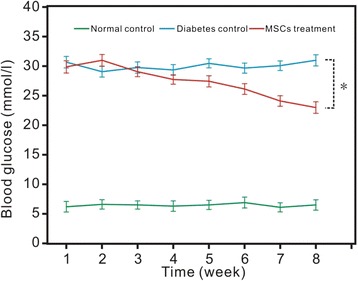
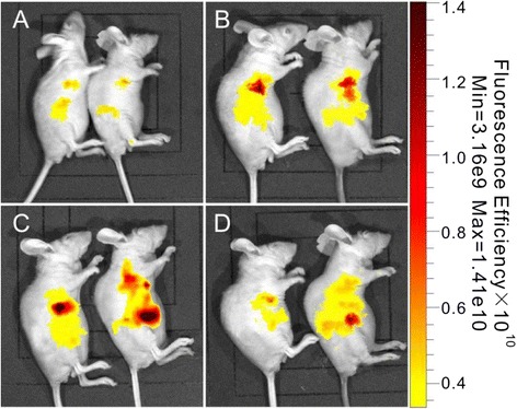
Mesenchymal stem cells (MSCs) have been used for therapy of type 1 diabetes mellitus. However, the in vivo distribution and therapeutic effects of transplanted MSCs are not clarified well. Herein, we reported that CdSe/ZnS quantum dots-labeled MSCs were prepared for targeted fluorescence imaging and therapy of pancreas tissues in rat models with type 1 diabetes. CdSe/ZnS quantum dots were synthesized, their biocompatibility was evaluated, and then, the appropriate concentration of quantum dots was selected to label MSCs. CdSe/ZnS quantum dots-labeled MSCs were injected into mouse models with type 1 diabetes via tail vessel and then were observed by using the Bruker In-Vivo F PRO system, and the blood glucose levels were monitored for 8 weeks. Results showed that prepared CdSe/ZnS quantum dots owned good biocompatibility. Significant differences existed in distribution of quantum dots-labeled MSCs between normal control rats and diabetic rats (p < 0.05). The ratios of the fluorescence intensity (RFI) analysis showed an accumulation rate of MSCs in the pancreas of rats in the diabetes group which was about 32 %, while that in the normal control group rats was about 18 %. The blood glucose levels were also monitored for 8 weeks after quantum dots-labeled MSC injection. Statistical differences existed between the blood glucose levels of the diabetic rat control group and MSC-injected diabetic rat group (p < 0.01), and the MSC-injected diabetic rat group displayed lower blood glucose levels. In conclusion, CdSe/ZnS-labeled MSCs can target in vivo pancreas tissues in diabetic rats, and significantly reduce the blood glucose levels in diabetic rats, and own potential application in therapy of diabetic patients in the near future.

摘要
https://cdn.ncbi.nlm.nih.gov/pmc/blobs/ea10/4469594/20439883c13b/11671_2015_959_Fig11_HTML.jpg
https://cdn.ncbi.nlm.nih.gov/pmc/blobs/ea10/4469594/0ba7f5e4ff31/11671_2015_959_Fig1_HTML.jpg
https://cdn.ncbi.nlm.nih.gov/pmc/blobs/ea10/4469594/d1b8b283d198/11671_2015_959_Fig2_HTML.jpg
https://cdn.ncbi.nlm.nih.gov/pmc/blobs/ea10/4469594/7ed7206acb71/11671_2015_959_Fig3_HTML.jpg
https://cdn.ncbi.nlm.nih.gov/pmc/blobs/ea10/4469594/65d0aaa1c2db/11671_2015_959_Fig4_HTML.jpg
https://cdn.ncbi.nlm.nih.gov/pmc/blobs/ea10/4469594/9f3c4a33c992/11671_2015_959_Fig5_HTML.jpg
https://cdn.ncbi.nlm.nih.gov/pmc/blobs/ea10/4469594/469fa98f0274/11671_2015_959_Fig6_HTML.jpg
https://cdn.ncbi.nlm.nih.gov/pmc/blobs/ea10/4469594/8029dfea6c7b/11671_2015_959_Fig7_HTML.jpg
https://cdn.ncbi.nlm.nih.gov/pmc/blobs/ea10/4469594/8bc281789b14/11671_2015_959_Fig8_HTML.jpg
https://cdn.ncbi.nlm.nih.gov/pmc/blobs/ea10/4469594/e83b460fa79c/11671_2015_959_Fig9_HTML.jpg
https://cdn.ncbi.nlm.nih.gov/pmc/blobs/ea10/4469594/037bb911cb14/11671_2015_959_Fig10_HTML.jpg
https://cdn.ncbi.nlm.nih.gov/pmc/blobs/ea10/4469594/20439883c13b/11671_2015_959_Fig11_HTML.jpg

相似文献

1
CdSe/ZnS Quantum Dots-Labeled Mesenchymal Stem Cells for Targeted Fluorescence Imaging of Pancreas Tissues and Therapy of Type 1 Diabetic Rats.
Nanoscale Res Lett. 2015 Dec;10(1):959. doi: 10.1186/s11671-015-0959-3. Epub 2015 Jun 13.
2
Allogeneic diabetic mesenchymal stem cells transplantation in streptozotocin-induced diabetic rat.链脲佐菌素诱导的糖尿病大鼠同种异体糖尿病间充质干细胞移植
Clin Invest Med. 2008 Dec 1;31(6):E328-37. doi: 10.25011/cim.v31i6.4918.
3
Thiolate-Capped CdSe/ZnS Core-Shell Quantum Dots for the Sensitive Detection of Glucose.巯基乙酸盐封端的 CdSe/ZnS 核壳量子点用于葡萄糖的灵敏检测。
Sensors (Basel). 2017 Jul 1;17(7):1537. doi: 10.3390/s17071537.
4
Examination of the stability of hydrophobic (CdSe)ZnS quantum dots in the digestive tract of rats.大鼠消化道中疏水性(CdSe)ZnS量子点的稳定性研究。
Photochem Photobiol Sci. 2008 Jun;7(6):725-9. doi: 10.1039/b707920f. Epub 2008 Apr 18.
5
Biocompatibility of quantum dots (CdSe/ZnS ) in human amniotic membrane-derived mesenchymal stem cells in vitro.量子点(CdSe/ZnS)在人羊膜间充质干细胞中的体外生物相容性
Biomed Pap Med Fac Univ Palacky Olomouc Czech Repub. 2015 Jun;159(2):227-33. doi: 10.5507/bp.2014.045. Epub 2014 Oct 2.
6
Applications of mesenchymal stem cells labeled with Tat peptide conjugated quantum dots to cell tracking in mouse body.用Tat肽偶联量子点标记的间充质干细胞在小鼠体内细胞追踪中的应用。
Bioconjug Chem. 2008 Feb;19(2):421-7. doi: 10.1021/bc0700685. Epub 2007 Dec 15.
7
Quantum dots labelling allows detection of the homing of mesenchymal stem cells administered as immunomodulatory therapy in an experimental model of pancreatic islets transplantation.量子点标记能够在胰岛移植实验模型中检测作为免疫调节疗法施用的间充质干细胞的归巢情况。
J Anat. 2017 Mar;230(3):381-388. doi: 10.1111/joa.12563. Epub 2016 Nov 15.
8
Mesenchymal stem cells and differentiated insulin producing cells are new horizons for pancreatic regeneration in type I diabetes mellitus.间充质干细胞和分化的胰岛素产生细胞是I型糖尿病胰腺再生的新方向。
Int J Biochem Cell Biol. 2017 Jun;87:77-85. doi: 10.1016/j.biocel.2017.03.018. Epub 2017 Apr 3.
9
CdSe@ZnS/ZnS quantum dots loaded in polymeric micelles as a pH-triggerable targeting fluorescence imaging probe for detecting cerebral ischemic area.负载于聚合物胶束中的CdSe@ZnS/ZnS量子点作为一种pH触发的靶向荧光成像探针用于检测脑缺血区域。
Colloids Surf B Biointerfaces. 2017 Jul 1;155:497-506. doi: 10.1016/j.colsurfb.2017.04.054. Epub 2017 Apr 27.
10
CdSe/ZnS quantum dots induce photodynamic effects and cytotoxicity in pancreatic cancer cells.硒化镉/硫化锌量子点诱导胰腺癌细胞产生光动力效应和细胞毒性。
World J Gastroenterol. 2016 Jun 7;22(21):5012-22. doi: 10.3748/wjg.v22.i21.5012.

引用本文的文献

1
Angiotensin 1 peptide-conjugated CdSe/ZnS quantum dots for cardiac-specific hydrogen sulfide targeted therapy in myocardial ischemia-reperfusion injury.血管紧张素1肽偶联的CdSe/ZnS量子点用于心肌缺血再灌注损伤的心脏特异性硫化氢靶向治疗
Front Pharmacol. 2024 Oct 2;15:1435282. doi: 10.3389/fphar.2024.1435282. eCollection 2024.
2
Precision Nanomedicine with Bio-Inspired Nanosystems: Recent Trends and Challenges in Mesenchymal Stem Cells Membrane-Coated Bioengineered Nanocarriers in Targeted Nanotherapeutics.具有生物启发纳米系统的精准纳米医学:靶向纳米治疗中基于间充质干细胞膜包覆生物工程纳米载体的最新趋势与挑战
J Xenobiot. 2024 Jun 24;14(3):827-872. doi: 10.3390/jox14030047.
3

本文引用的文献

1
In vivo real-time visualization of mesenchymal stem cells tropism for cutaneous regeneration using NIR-II fluorescence imaging.利用近红外二区荧光成像技术在体实时可视化间充质干细胞对皮肤再生的趋向性。
Biomaterials. 2015 Jun;53:265-73. doi: 10.1016/j.biomaterials.2015.02.090. Epub 2015 Mar 16.
2
Cation-inverting-injection: a novel method for synthesis of aqueous ZnSe quantum dots with bright excitionic emission and suppressed trap emission.阳离子反转注入法:一种合成具有明亮激子发射和抑制陷阱发射的水溶性硒化锌量子点的新方法。
Nanotechnology. 2014 Jul 25;25(29):295602. doi: 10.1088/0957-4484/25/29/295602. Epub 2014 Jul 1.
3
Real-Time Quantum Dots Imaging to Track Transplanted Adipose Stem Cells in Different Inflammatory States of Acute Liver Failure Mice.
实时量子点成像技术追踪急性肝衰竭小鼠不同炎症状态下移植的脂肪干细胞。
Cell Transplant. 2023 Jan-Dec;32:9636897231176442. doi: 10.1177/09636897231176442.
4
Advanced Nanotechnology Approaches as Emerging Tools in Cellular-Based Technologies.先进的纳米技术方法作为基于细胞的技术中的新兴工具。
Adv Exp Med Biol. 2023;1409:127-144. doi: 10.1007/5584_2022_725.
5
Designing inorganic nanomaterials for vaccines and immunotherapies.设计用于疫苗和免疫疗法的无机纳米材料。
Nano Today. 2019 Aug;27:73-98. doi: 10.1016/j.nantod.2019.04.005. Epub 2019 May 29.
6
A brief review of cytotoxicity of nanoparticles on mesenchymal stem cells in regenerative medicine.纳米颗粒对再生医学中间充质干细胞细胞毒性的简要综述。
Int J Nanomedicine. 2019 May 24;14:3875-3892. doi: 10.2147/IJN.S205574. eCollection 2019.
7
Targeted migration of bone marrow mesenchymal stem cells inhibits silica-induced pulmonary fibrosis in rats.骨髓间充质干细胞靶向迁移抑制大鼠二氧化硅诱导的肺纤维化。
Stem Cell Res Ther. 2018 Dec 4;9(1):335. doi: 10.1186/s13287-018-1083-y.
8
Cytocompatible and multifunctional polymeric nanoparticles for transportation of bioactive molecules into and within cells.用于将生物活性分子运输到细胞内和细胞中的具有细胞相容性和多功能的聚合物纳米颗粒。
Sci Technol Adv Mater. 2016 Jul 6;17(1):300-312. doi: 10.1080/14686996.2016.1190257. eCollection 2016.
BRCAA1 antibody- and Her2 antibody-conjugated amphiphilic polymer engineered CdSe/ZnS quantum dots for targeted imaging of gastric cancer.
BRCAA1 抗体和 Her2 抗体偶联的两亲聚合物修饰的 CdSe/ZnS 量子点用于胃癌的靶向成像。
Nanoscale Res Lett. 2014 May 19;9(1):244. doi: 10.1186/1556-276X-9-244. eCollection 2014.
4
Mesenchymal stem cells: new players in retinopathy therapy.间充质干细胞:视网膜病变治疗中的新角色。
Front Endocrinol (Lausanne). 2014 Apr 24;5:59. doi: 10.3389/fendo.2014.00059. eCollection 2014.
5
Cell replacement strategies aimed at reconstitution of the β-cell compartment in type 1 diabetes.旨在重建1型糖尿病β细胞区室的细胞替代策略。
Diabetes. 2014 May;63(5):1433-44. doi: 10.2337/db13-1742.
6
Bone marrow mesenchymal stem cells promote the repair of islets from diabetic mice through paracrine actions.骨髓间充质干细胞通过旁分泌作用促进糖尿病小鼠胰岛的修复。
Mol Cell Endocrinol. 2014 May 5;388(1-2):41-50. doi: 10.1016/j.mce.2014.03.004. Epub 2014 Mar 22.
7
Effect and mechanisms of human Wharton's jelly-derived mesenchymal stem cells on type 1 diabetes in NOD model.人牙髓间充质干细胞对 NOD 模型 1 型糖尿病的作用及其机制。
Endocrine. 2015 Feb;48(1):124-34. doi: 10.1007/s12020-014-0219-9. Epub 2014 Mar 4.
8
Quantitative metabolic imaging using endogenous fluorescence to detect stem cell differentiation.利用内源性荧光进行定量代谢成像以检测干细胞分化。
Sci Rep. 2013 Dec 5;3:3432. doi: 10.1038/srep03432.
9
In vivo real-time visualization of tissue blood flow and angiogenesis using Ag2S quantum dots in the NIR-II window.利用近红外二区的 Ag2S 量子点对组织血流和血管生成进行体内实时可视化。
Biomaterials. 2014 Jan;35(1):393-400. doi: 10.1016/j.biomaterials.2013.10.010. Epub 2013 Oct 15.
10
Mesenchymal stem cell therapy in diabetes mellitus: progress and challenges.间充质干细胞疗法治疗糖尿病:进展与挑战。
J Nucleic Acids. 2013;2013:194858. doi: 10.1155/2013/194858. Epub 2013 May 15.