• 文献检索
  • 文档翻译
  • 深度研究
  • 学术资讯
  • Suppr Zotero 插件Zotero 插件
  • 邀请有礼
  • 套餐&价格
  • 历史记录
应用&插件
Suppr Zotero 插件Zotero 插件浏览器插件Mac 客户端Windows 客户端微信小程序
定价
高级版会员购买积分包购买API积分包
服务
文献检索文档翻译深度研究API 文档MCP 服务
关于我们
关于 Suppr公司介绍联系我们用户协议隐私条款
关注我们

Suppr 超能文献

核心技术专利:CN118964589B侵权必究
粤ICP备2023148730 号-1Suppr @ 2026

文献检索

告别复杂PubMed语法,用中文像聊天一样搜索,搜遍4000万医学文献。AI智能推荐,让科研检索更轻松。

立即免费搜索

文件翻译

保留排版,准确专业,支持PDF/Word/PPT等文件格式,支持 12+语言互译。

免费翻译文档

深度研究

AI帮你快速写综述,25分钟生成高质量综述,智能提取关键信息,辅助科研写作。

立即免费体验

基于自适应Sigmoid函数和空间可变颜色再现的内镜图像颜色增强

Color Enhancement in Endoscopic Images Using Adaptive Sigmoid Function and Space Variant Color Reproduction.

作者信息

Imtiaz Mohammad S, Wahid Khan A

机构信息

Department of Electrical and Computer Engineering, University of Saskatchewan, Saskatoon, SK, Canada S7N 5A9.

出版信息

Comput Math Methods Med. 2015;2015:607407. doi: 10.1155/2015/607407. Epub 2015 May 18.

DOI:10.1155/2015/607407
PMID:26089969
原文链接:https://pmc.ncbi.nlm.nih.gov/articles/PMC4450341/
Abstract

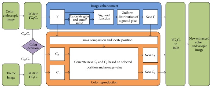
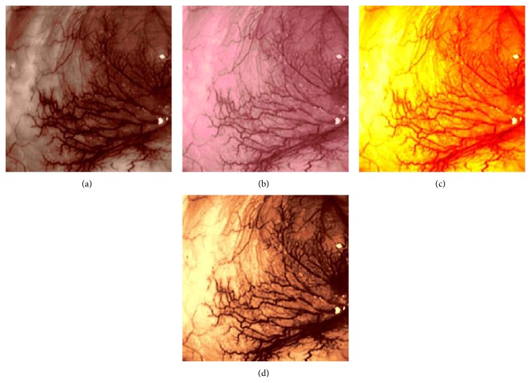
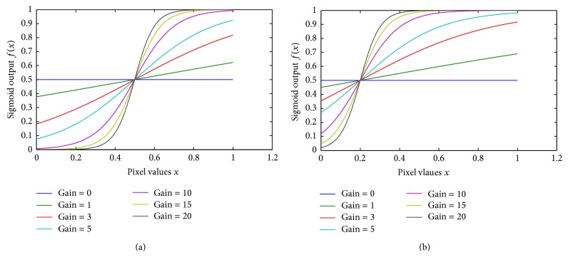
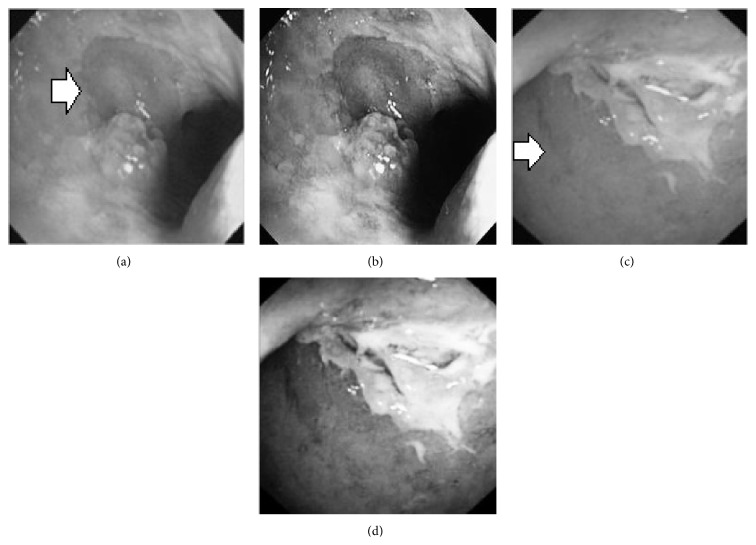
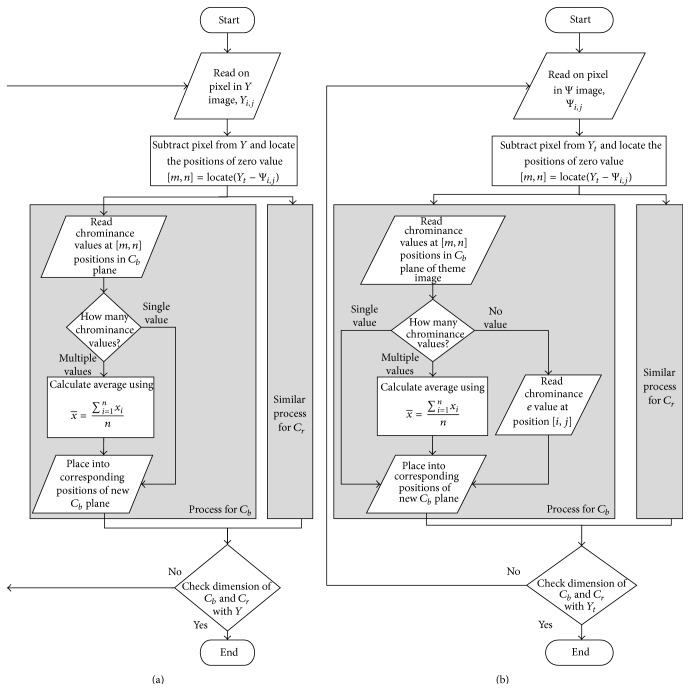
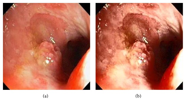
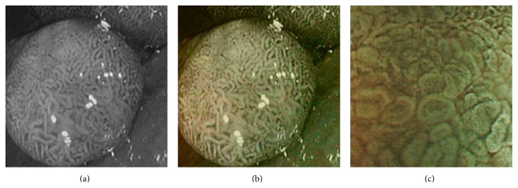
Modern endoscopes play an important role in diagnosing various gastrointestinal (GI) tract related diseases. The improved visual quality of endoscopic images can provide better diagnosis. This paper presents an efficient color image enhancement method for endoscopic images. It is achieved in two stages: image enhancement at gray level followed by space variant chrominance mapping color reproduction. Image enhancement is achieved by performing adaptive sigmoid function and uniform distribution of sigmoid pixels. Secondly, a space variant chrominance mapping color reproduction is used to generate new chrominance components. The proposed method is used on low contrast color white light images (WLI) to enhance and highlight the vascular and mucosa structures of the GI tract. The method is also used to colorize grayscale narrow band images (NBI) and video frames. The focus value and color enhancement factor show that the enhancement level in the processed image is greatly increased compared to the original endoscopic image. The overall contrast level of the processed image is higher than the original image. The color similarity test has proved that the proposed method does not add any additional color which is not present in the original image. The algorithm has low complexity with an execution speed faster than other related methods.

摘要

现代内窥镜在诊断各种胃肠道相关疾病中发挥着重要作用。内窥镜图像视觉质量的提高有助于更好地进行诊断。本文提出了一种针对内窥镜图像的高效彩色图像增强方法。该方法分两个阶段实现:灰度级图像增强,随后是空间可变色度映射颜色再现。图像增强通过执行自适应Sigmoid函数和Sigmoid像素的均匀分布来实现。其次,使用空间可变色度映射颜色再现来生成新的色度分量。所提出的方法用于低对比度彩色白光图像(WLI),以增强和突出胃肠道的血管和黏膜结构。该方法还用于对灰度窄带图像(NBI)和视频帧进行彩色化处理。聚焦值和颜色增强因子表明,与原始内窥镜图像相比,处理后的图像增强水平有了显著提高。处理后图像的整体对比度水平高于原始图像。颜色相似性测试证明,所提出的方法不会添加原始图像中不存在的任何额外颜色。该算法复杂度低,执行速度比其他相关方法更快。

https://cdn.ncbi.nlm.nih.gov/pmc/blobs/ef90/4450341/e1aacdabf11d/CMMM2015-607407.007.jpg
https://cdn.ncbi.nlm.nih.gov/pmc/blobs/ef90/4450341/ac4554c876b9/CMMM2015-607407.001.jpg
https://cdn.ncbi.nlm.nih.gov/pmc/blobs/ef90/4450341/fcbe6bae77af/CMMM2015-607407.002.jpg
https://cdn.ncbi.nlm.nih.gov/pmc/blobs/ef90/4450341/bb4db022378f/CMMM2015-607407.003.jpg
https://cdn.ncbi.nlm.nih.gov/pmc/blobs/ef90/4450341/2f0eb88b2b1e/CMMM2015-607407.004.jpg
https://cdn.ncbi.nlm.nih.gov/pmc/blobs/ef90/4450341/5cd6bf3782dd/CMMM2015-607407.005.jpg
https://cdn.ncbi.nlm.nih.gov/pmc/blobs/ef90/4450341/ef7fd9fd9c10/CMMM2015-607407.006.jpg
https://cdn.ncbi.nlm.nih.gov/pmc/blobs/ef90/4450341/e1aacdabf11d/CMMM2015-607407.007.jpg

相似文献

1
Color Enhancement in Endoscopic Images Using Adaptive Sigmoid Function and Space Variant Color Reproduction.基于自适应Sigmoid函数和空间可变颜色再现的内镜图像颜色增强
Comput Math Methods Med. 2015;2015:607407. doi: 10.1155/2015/607407. Epub 2015 May 18.
2
Tri-Scan: A Three Stage Color Enhancement Tool for Endoscopic Images.Tri-Scan:一种用于内镜图像的三阶段色彩增强工具。
J Med Syst. 2017 Jun;41(6):102. doi: 10.1007/s10916-017-0738-z. Epub 2017 May 20.
3
Image enhancement and space-variant color reproduction method for endoscopic images using adaptive sigmoid function.基于自适应Sigmoid函数的内镜图像增强与空间可变色彩再现方法
Annu Int Conf IEEE Eng Med Biol Soc. 2014;2014:3905-8. doi: 10.1109/EMBC.2014.6944477.
4
Color reproduction and processing algorithm based on real-time mapping for endoscopic images.基于实时映射的内镜图像色彩再现与处理算法
Springerplus. 2016 Jan 6;5:17. doi: 10.1186/s40064-015-1612-4. eCollection 2016.
5
Wireless Micro-Ball endoscopic image enhancement using histogram information.
Annu Int Conf IEEE Eng Med Biol Soc. 2014;2014:3337-40. doi: 10.1109/EMBC.2014.6944337.
6
Automatic detection of informative frames from wireless capsule endoscopy images.无线胶囊内窥镜图像中信息帧的自动检测。
Med Image Anal. 2010 Jun;14(3):449-70. doi: 10.1016/j.media.2009.12.001. Epub 2010 Jan 4.
7
Wireless capsule endoscopy images enhancement by tensor based diffusion.
Conf Proc IEEE Eng Med Biol Soc. 2006;2006:4861-4. doi: 10.1109/IEMBS.2006.260002.
8
Adaptive Image Enhancement Based on Guide Image and Fraction-Power Transformation for Wireless Capsule Endoscopy.基于引导图像和分数幂变换的无线胶囊内窥镜自适应图像增强。
IEEE Trans Biomed Circuits Syst. 2018 Oct;12(5):993-1003. doi: 10.1109/TBCAS.2018.2869530.
9
Deep unsupervised endoscopic image enhancement based on multi-image fusion.基于多图像融合的深度无监督内窥镜图像增强。
Comput Methods Programs Biomed. 2022 Jun;221:106800. doi: 10.1016/j.cmpb.2022.106800. Epub 2022 Apr 26.
10
Image partitioning and illumination in image-based pose detection for teleoperated flexible endoscopes.基于图像的远程操作柔性内窥镜位姿检测中的图像分区和光照。
Artif Intell Med. 2013 Nov;59(3):185-96. doi: 10.1016/j.artmed.2013.09.002. Epub 2013 Oct 10.

引用本文的文献

1
A -Extension of Sigmoid Functions and the Application for Enhancement of Ultrasound Images.A - 西格蒙德函数的扩展及其在超声图像增强中的应用。
Entropy (Basel). 2019 Apr 23;21(4):430. doi: 10.3390/e21040430.
2
Evaluation of Reconstruction Parameters for 2-D Comb-Push Ultrasound Shear Wave Elastography.二维梳状激励超声剪切波弹性成像重建参数的评估。
IEEE Trans Ultrason Ferroelectr Freq Control. 2019 Feb;66(2):254-263. doi: 10.1109/TUFFC.2018.2884348. Epub 2018 Nov 30.
3
CHOBS: Color Histogram of Block Statistics for Automatic Bleeding Detection in Wireless Capsule Endoscopy Video.

本文引用的文献

1
Image enhancement and space-variant color reproduction method for endoscopic images using adaptive sigmoid function.基于自适应Sigmoid函数的内镜图像增强与空间可变色彩再现方法
Annu Int Conf IEEE Eng Med Biol Soc. 2014;2014:3905-8. doi: 10.1109/EMBC.2014.6944477.
2
A wireless narrowband imaging chip for capsule endoscope.一种用于胶囊内窥镜的无线窄带成像芯片。
IEEE Trans Biomed Circuits Syst. 2010 Dec;4(6):462-8. doi: 10.1109/TBCAS.2010.2079932.
3
Comparison of detection and miss rates of narrow band imaging, flexible spectral imaging chromoendoscopy and white light at screening colonoscopy: a randomised controlled back-to-back study.
CHOBS:无线胶囊内镜视频中用于自动出血检测的块统计颜色直方图
IEEE J Transl Eng Health Med. 2018 Jan 24;6:1800112. doi: 10.1109/JTEHM.2017.2756034. eCollection 2018.
4
Automatic deformable PET/MRI registration for preclinical studies based on B-splines and non-linear intensity transformation.基于 B 样条和非线性强度变换的小动物 PET/MRI 自动配准。
Med Biol Eng Comput. 2018 Sep;56(9):1531-1539. doi: 10.1007/s11517-018-1797-0. Epub 2018 Feb 7.
5
Advanced Image Enhancement Method for Distant Vessels and Structures in Capsule Endoscopy.胶囊内镜中远侧血管和结构的高级图像增强方法。
Comput Math Methods Med. 2017;2017:9813165. doi: 10.1155/2017/9813165. Epub 2017 Oct 31.
6
Frontiers of robotic endoscopic capsules: a review.机器人内镜胶囊前沿:综述
J Microbio Robot. 2016;11(1):1-18. doi: 10.1007/s12213-016-0087-x. Epub 2016 May 2.
窄带成像、弹性光谱成像 chromoendoscopy 和白光在筛查结肠镜检查中的检测和漏诊率比较:一项随机对照背靠背研究。
Gut. 2014 May;63(5):785-91. doi: 10.1136/gutjnl-2013-304578. Epub 2013 Jul 12.
4
Robust automatic focus algorithm for low contrast images using a new contrast measure.使用新对比度度量的用于低对比度图像的鲁棒自动对焦算法。
Sensors (Basel). 2011;11(9):8281-94. doi: 10.3390/s110908281. Epub 2011 Aug 25.
5
Autofluorescence imaging endoscopy for screening of esophageal squamous mucosal high-grade neoplasia: a phase II study.荧光内镜自体荧光成像筛查食管鳞状上皮高级别瘤变:Ⅱ期研究。
J Gastroenterol Hepatol. 2012 Jan;27(1):86-90. doi: 10.1111/j.1440-1746.2011.06850.x.
6
A summary-statistic representation in peripheral vision explains visual crowding.外周视觉中的汇总统计表示解释了视觉拥挤现象。
J Vis. 2009 Nov 19;9(12):13.1-18. doi: 10.1167/9.12.13.
7
Fundus autofluorescence imaging: review and perspectives.眼底自发荧光成像:综述与展望。
Retina. 2008 Mar;28(3):385-409. doi: 10.1097/IAE.0b013e318164a907.
8
Chromoendoscopy and narrow-band imaging compared with high-resolution magnification endoscopy in Barrett's esophagus.巴雷特食管中色素内镜检查和窄带成像与高分辨率放大内镜检查的比较
Gastroenterology. 2008 Mar;134(3):670-9. doi: 10.1053/j.gastro.2008.01.003. Epub 2008 Jan 10.
9
American Gastroenterological Association (AGA) Institute technology assessment on image-enhanced endoscopy.美国胃肠病学会(AGA)研究所关于图像增强内镜检查的技术评估
Gastroenterology. 2008 Jan;134(1):327-40. doi: 10.1053/j.gastro.2007.10.062. Epub 2007 Oct 30.
10
Computed virtual chromoendoscopy: a new tool for enhancing tissue surface structures.计算机虚拟染色内镜检查:一种增强组织表面结构的新工具。
Endoscopy. 2007 Jan;39(1):80-3. doi: 10.1055/s-2006-945045.