Guizard Nicolas, Coupé Pierrick, Fonov Vladimir S, Manjón Jose V, Arnold Douglas L, Collins D Louis
Montreal Neurological Institute, McGill University, Canada.
Laboratoire Bordelais de Recherche en Informatique, Unité Mixte de Recherche CNRS (UMR 5800), PICTURA Research Group, 351, Talence, France.
Neuroimage Clin. 2015 May 13;8:376-89. doi: 10.1016/j.nicl.2015.05.001. eCollection 2015.
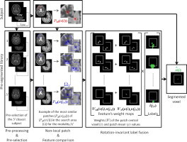
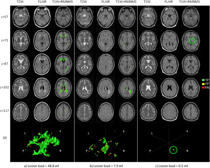
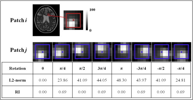
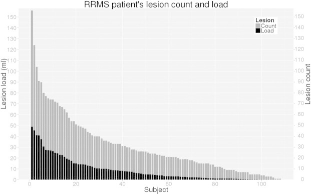
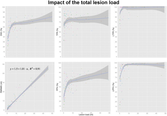
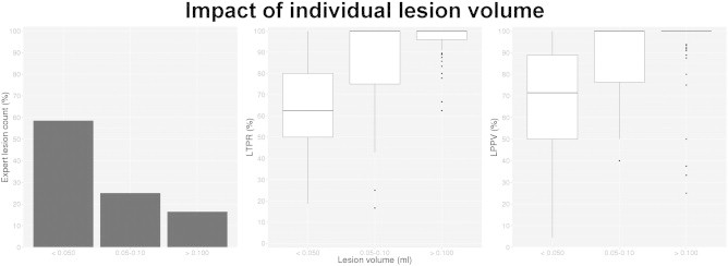
Multiple sclerosis (MS) lesion segmentation is crucial for evaluating disease burden, determining disease progression and measuring the impact of new clinical treatments. MS lesions can vary in size, location and intensity, making automatic segmentation challenging. In this paper, we propose a new supervised method to segment MS lesions from 3D magnetic resonance (MR) images using non-local means (NLM). The method uses a multi-channel and rotation-invariant distance measure to account for the diversity of MS lesions. The proposed segmentation method, rotation-invariant multi-contrast non-local means segmentation (RMNMS), captures the MS lesion spatial distribution and can accurately and robustly identify lesions regardless of their orientation, shape or size. An internal validation on a large clinical magnetic resonance imaging (MRI) dataset of MS patients demonstrated a good similarity measure result (Dice similarity = 60.1% and sensitivity = 75.4%), a strong correlation between expert and automatic lesion load volumes (R(2) = 0.91), and a strong ability to detect lesions of different sizes and in varying spatial locations (lesion detection rate = 79.8%). On the independent MS Grand Challenge (MSGC) dataset validation, our method provided competitive results with state-of-the-art supervised and unsupervised methods. Qualitative visual and quantitative voxel- and lesion-wise evaluations demonstrated the accuracy of RMNMS method.
多发性硬化症(MS)病灶分割对于评估疾病负担、确定疾病进展以及衡量新临床治疗的效果至关重要。MS病灶在大小、位置和强度上存在差异,这使得自动分割具有挑战性。在本文中,我们提出了一种新的监督方法,使用非局部均值(NLM)从三维磁共振(MR)图像中分割MS病灶。该方法使用多通道和旋转不变距离度量来考虑MS病灶的多样性。所提出的分割方法,即旋转不变多对比度非局部均值分割(RMNMS),能够捕捉MS病灶的空间分布,并且无论病灶的方向、形状或大小如何,都能准确且稳健地识别病灶。对大量MS患者的临床磁共振成像(MRI)数据集进行的内部验证表明,相似性度量结果良好(骰子相似系数 = 60.1%,灵敏度 = 75.4%),专家手动测量的病灶负荷体积与自动分割结果之间具有很强的相关性(R(2) = 0.91),并且具有很强的检测不同大小和不同空间位置病灶的能力(病灶检测率 = 79.8%)。在独立的MS大挑战(MSGC)数据集验证中,我们的方法与最先进的监督和无监督方法相比提供了具有竞争力的结果。定性的视觉评估以及定量体素级和病灶级评估都证明了RMNMS方法的准确性。